Light instead of radiation: seeing an organ without a needle express
Radioactive tracers are like a courier with a beacon: you inject it into the body, and it highlights the target organ on the scan. But this courier comes with strict rules: you can't send it often, it quickly 'gets tired,' and it costs a lot. Doctors have long dreamed of a way to see an organ without radiation.
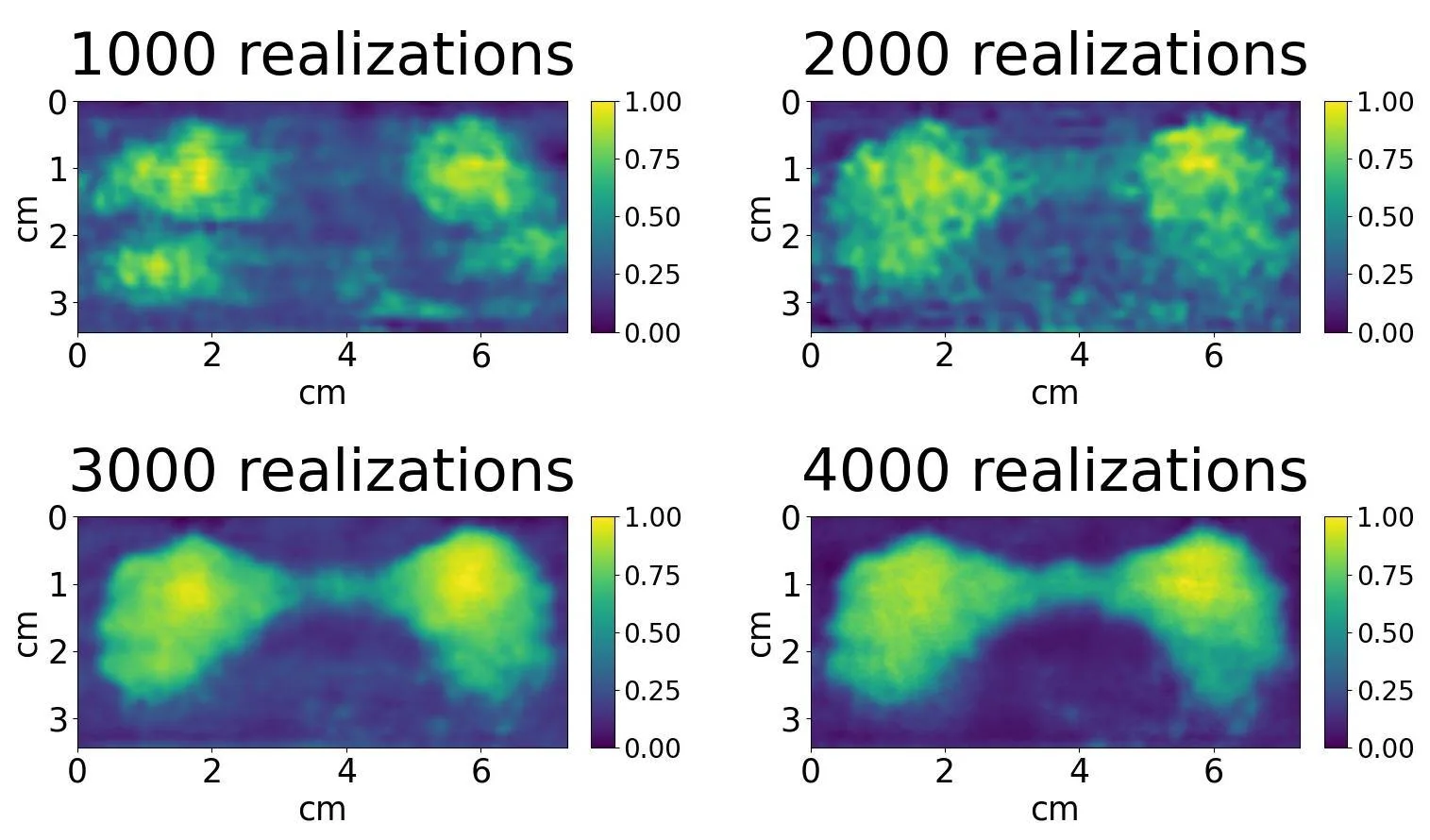
New work shows that you can illuminate an organ with ordinary X-rays, and iodine atoms will do the responding: they flash with secondary emission, like fireflies answering a flashlight. A detector captures the light from iodine and assembles an image from these flashes. In the experiment, they took a plastic model of the thyroid gland, filled it with an iodine solution, and obtained an image with clarity close to computed tomography.
The trick of the method lies in how light is used. Instead of a continuous beam, the X-rays are directed at the organ as a pattern of many small spots, as if a flashlight shone through a perforated stencil. The computer knows this pattern and, from how iodine responds from different points, reconstructs the full picture. This technique is called 'ghost imaging': the image is assembled from responses to known light patterns.
The main win is safety. Radioactive tracers cannot be injected often, but X-ray illumination can be repeated. This opens the door to regular thyroid checks for children and pregnant women, for whom radioactive methods are often contraindicated. It also removes the dependence on complex logistics: no need to transport short-lived isotopes to remote hospitals.
The thyroid gland is the only organ that actively accumulates iodine, so it can be 'lit up' even without a radioactive tracer, simply by giving a person ordinary iodine.