Tracking Prostate Cancer Metastases in Scans express
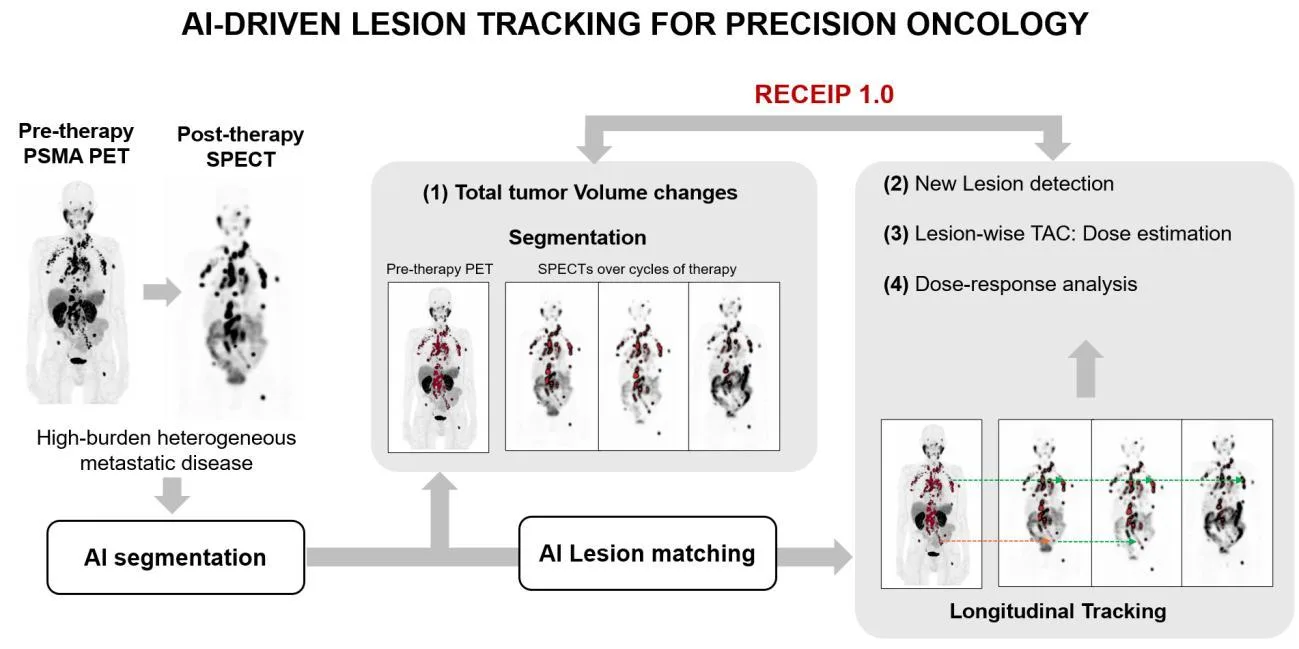
When treating prostate cancer, doctors often see not one tumor but many lesions in bones and lymph nodes. Each lesion can respond to therapy differently: one shrinks, another stays the same, a third grows. Previously, the disease was assessed as a whole, like judging the weather by the average temperature over a year. Now it's possible to track each lesion individually.
This is done using special scans: PET/CT with a substance that attaches to prostate cancer cells, and SPECT/CT after administering a therapeutic radioactive drug. A computer program identifies all suspicious areas on the scans and then matches them across different examinations, like an air traffic controller tracking each plane from takeoff to landing.
This approach helps doctors notice in time when a tumor stops responding to treatment and adjust their strategy. It also allows for more accurate radiation dose calculation for each lesion, sparing healthy tissue. Ultimately, treatment becomes more personal: not "against cancer in general," but against specific lesions in a specific person.
Original abstract
as written by the authors arXiv:2603.22666
Quantitative molecular imaging is central to treatment response assessment in oncology, yet clinical practice remains largely dominated by patient-level or limited target-lesion criteria that ignore inter-lesion heterogeneity. This limitation is particularly important in prostate cancer, where PSMA PET/CT can reveal extensive skeletal and nodal metastatic disease that often evolves heterogeneously under therapy. Accurate and scalable lesion segmentation and tracking across serial PSMA PET/CT and post-therapy SPECT/CT scans is therefore essential for implementing emerging PSMA-specific response frameworks, such as RECIP 1.0, and for enabling lesion-level dosimetry in 177Lu-PSMA radiopharmaceutical therapies (RPTs). This article examines clinical motivations, technical foundations, and future pathways for automated lesion tracking in prostate cancer imaging. We focus on the unique requirements introduced by PSMA PET/CT compared with FDG PET/CT and highlight the critical role of quantitative SPECT/CT in linking imaging-derived disease characterization with delivered therapeutic dose. Recent advances in AI-based segmentation and automated lesion matching now make scalable longitudinal lesion correspondence feasible, providing comprehensive infrastructure for standardized response assessment and personalized PSMA-based theranostics
PET/CT and SPECT/CT are like two sides of the same coin: the first scan shows where the cancer is hiding, and the second shows how much drug has reached it.
In Star Trek, the medical tricorder scans the body and instantly finds all problems. Similar algorithms are gradually entering real-world oncology.