Why Your Hand Trembles but Your Legs Won't Move express
For one person, the disease begins with a tremor in the hand — the cup dances on its saucer as they carry it to the table. For another, the legs suddenly refuse to move smoothly: the step becomes short and shuffling, as if the floor were suddenly covered in a sticky layer. These are not two different diseases but two faces of the same one — Parkinson's disease. But treatments and prognoses can differ, so doctors need to figure out as early as possible which face they're dealing with.
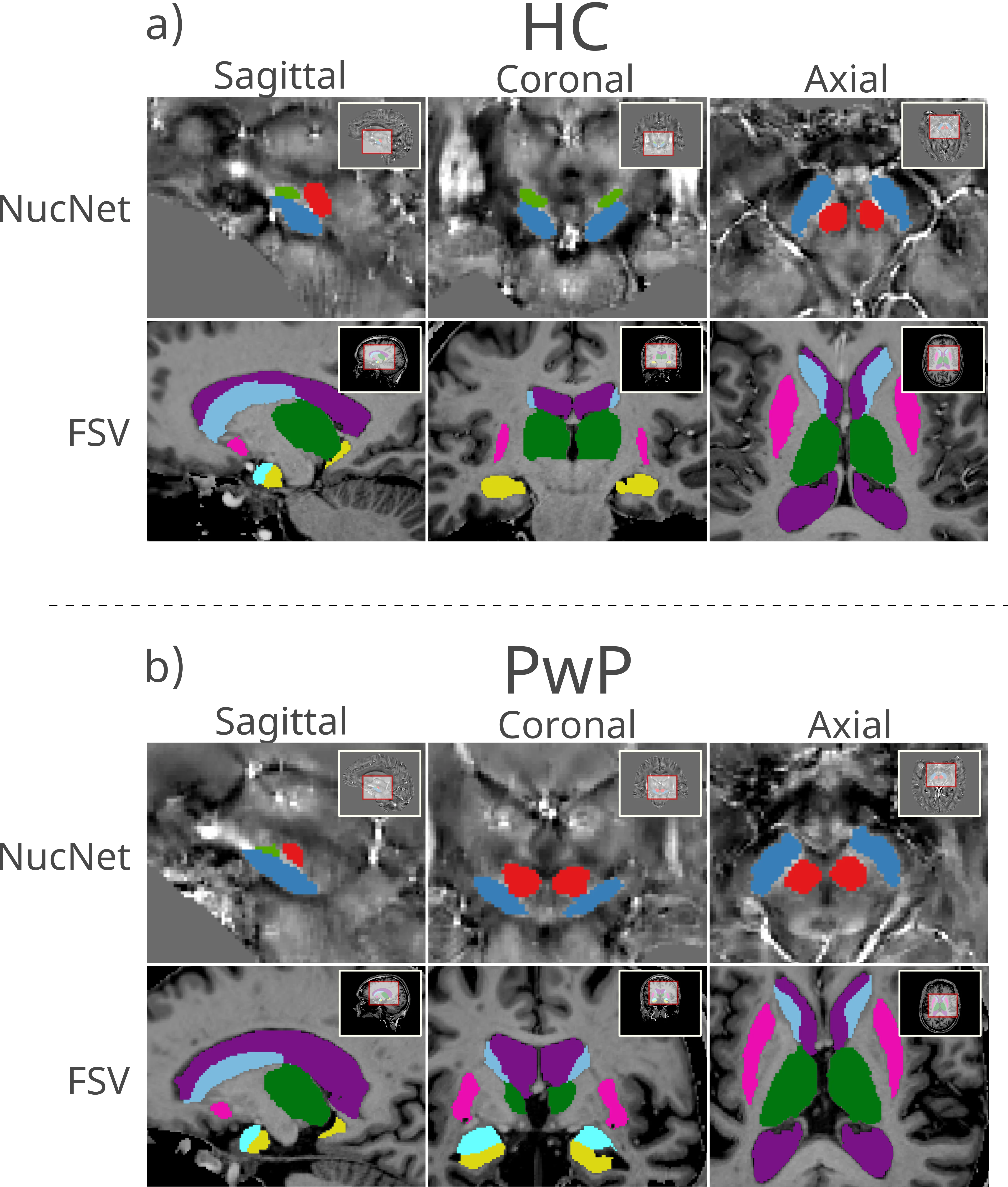
Researchers from the Netherlands let a computer look at the brains of 45 people—24 with Parkinson's and 21 healthy — using an ultra-powerful magnetic resonance imaging scanner with a field of 7 Tesla. For comparison, a typical hospital machine runs at 1.5 or 3 Tesla. At 7 Tesla, the images are so detailed that you can make out tiny structures deep inside the brain, where the early traces of the disease hide. A U-Net neural network was trained to automatically outline dozens of different zones in these images, like a child tracing outlines in a coloring book, but with precision down to fractions of a millimeter.
Next, from each zone they extracted a slew of numbers: tissue density, iron content, water molecule mobility, and so on. The result was a long list, like a huge report card with hundreds of grades for each patient. You'd think the more data, the better. But when they fed the entire report card to a classifier, a program that looks for patterns, it performed only mediocrely: it distinguished sick from healthy with 69% accuracy and mixed up the disease types. So the researchers applied feature selection — a method that keeps only the most "telling" measurements and discards the noise. It's like looking only at math and physics grades instead of the full school report card with gym and music, to predict whether someone will become an engineer. That's exactly what they did: from hundreds of indicators, they picked a handful of key ones, and the picture sharpened dramatically.
The main takeaway isn't that the machine is smarter than the doctor, but that the disease leaves clear, measurable traces in the brain, and there aren't that many of them. Just a few numbers from a single scan can tell how the disease will likely progress — whether it will take the tremor path or the stiffness path. This is a step toward getting a hint right from the MRI suite, instead of long observations and trial-and-error with medications. That same cup on the saucer where it all began might soon no longer be the only way to know what's going on inside.
Original abstract
as written by the authors arXiv:2605.24179
Parkinson's disease (PD) is a highly heterogeneous disease, including which motor symptoms are dominating. Imaging biomarkers that support subtype stratification could also improve biological understanding and study design, and enable personalized treatment strategies. This study evaluates whether deep-learning based automatic brain segmentation, in addition to quantitative maps from 7 Tesla MRI, can highlight differences between Healthy Controls (HC), Postural Instability and Gait Difficulty (PIGD) and Tremor Dominant (TD), and subsequently be used for objective PD stratification. The performance of machine learning classifiers may be improved with feature selection. 21 HC, and 24 people with PD (PwP) were included. The U-Net training was assessed with DSC. Two classification approaches using 5-fold cross-validation were defined across three tasks: (1) HC vs PwP; (2) PIGD vs TD; (3) multiclass, HC vs PIGD vs TD. Approach A used all extracted features. Approach B found the optimal subset of features for the classification tasks. The U-Net achieved mean DSC of 0.86 for all ROIs during training. Approach A: Task 1 best accuracy of 0.69 and best AUC of 0.73. Task 2 accuracy 0.69, AUC 0.90. Task 3 accuracy 0.62, AUC 0.66. Approach B: Task 1 accuracy of 0.82 and AUC of 0.93. Task 2 accuracy 1.00, AUC 1.00. Task 3 accuracy 0.73, AUC 0.91. DL-based segmentation combined with qMRI feature selection improved classification relative to using all features, supporting the potential of interpretable, low-dimensional imaging signatures for PD diagnosis support and phenotype stratification. Larger, multi-site studies are warranted to assess generalizability and stability.
The magnetic field of a 7-Tesla scanner is 140,000 times stronger than Earth's magnetic field: it can yank a steel chair out of your hands if you forget it in the scan room.