Viruses Under the X-Ray Beam express
An ordinary microscope is powerless when you need to examine a single virus in detail. It's too small. But there's a tool that fires flashes billions of times brighter than sunlight — an X-ray free-electron laser. It's designed to 'photograph' a particle before the beam vaporizes it. That's how scientists studied the bacteriophage PR772, sprayed into the air.
Each image isn't a familiar picture but a pattern of spots: that's how X-rays scatter when they hit the atoms of the virus. To assemble a three-dimensional model from these patterns, the authors used rotational invariants — mathematical quantities that don't change when an object is rotated. It's like recognizing a person by their unchanging facial features, no matter the angle you look from. Comparing thousands of such 'fingerprints,' the program computed the virus's shape.
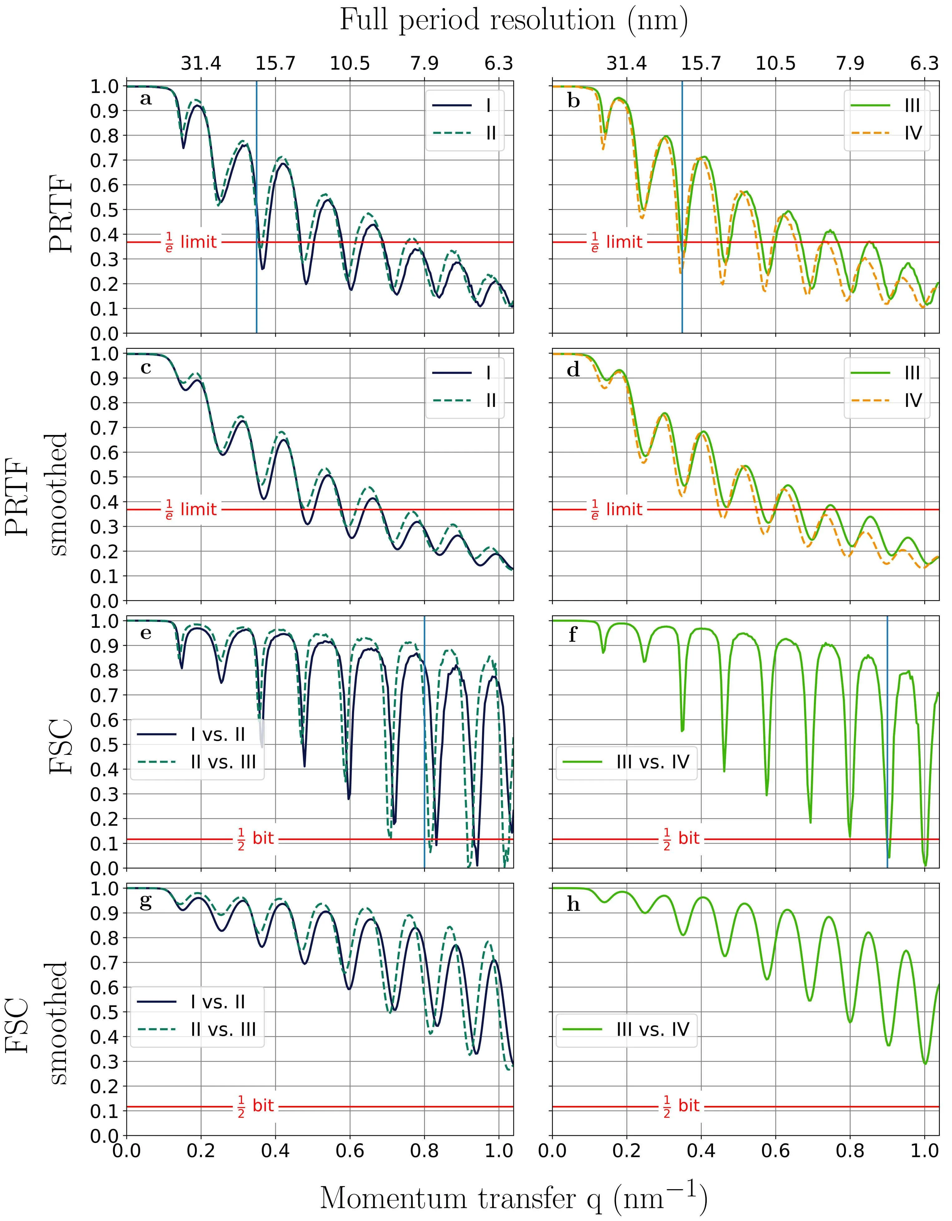
The image resolution was modest — about 20 nanometers (hundreds of times thinner than a human hair). But even so, the asymmetry of the shell and the inhomogeneity of the contents were visible. Previously, such details were lost in averaged models. Now the method allows tracking changes in viruses over time — for example, how they attach to a host cell.
An X-ray free-electron laser produces a flash lasting a few femtoseconds: that's like stretching one second to 30 million years and then taking a tiny fraction of that.
In Michael Crichton's novel 'The Andromeda Strain,' an alien virus is studied with an electron microscope and laser spectroscopy — much like in this work, except the laser didn't destroy the sample.