Microwaves Feel for the Edge of a Tumor express
A surgeon removes a breast tumor, but how do you know no cancer cells are left at the edge? Right now, the tissue goes to a lab, and the answer comes back days later. If the margin is not clean, a second surgery is needed. The new method promises to give the answer right in the operating room, while the patient is still on the table.
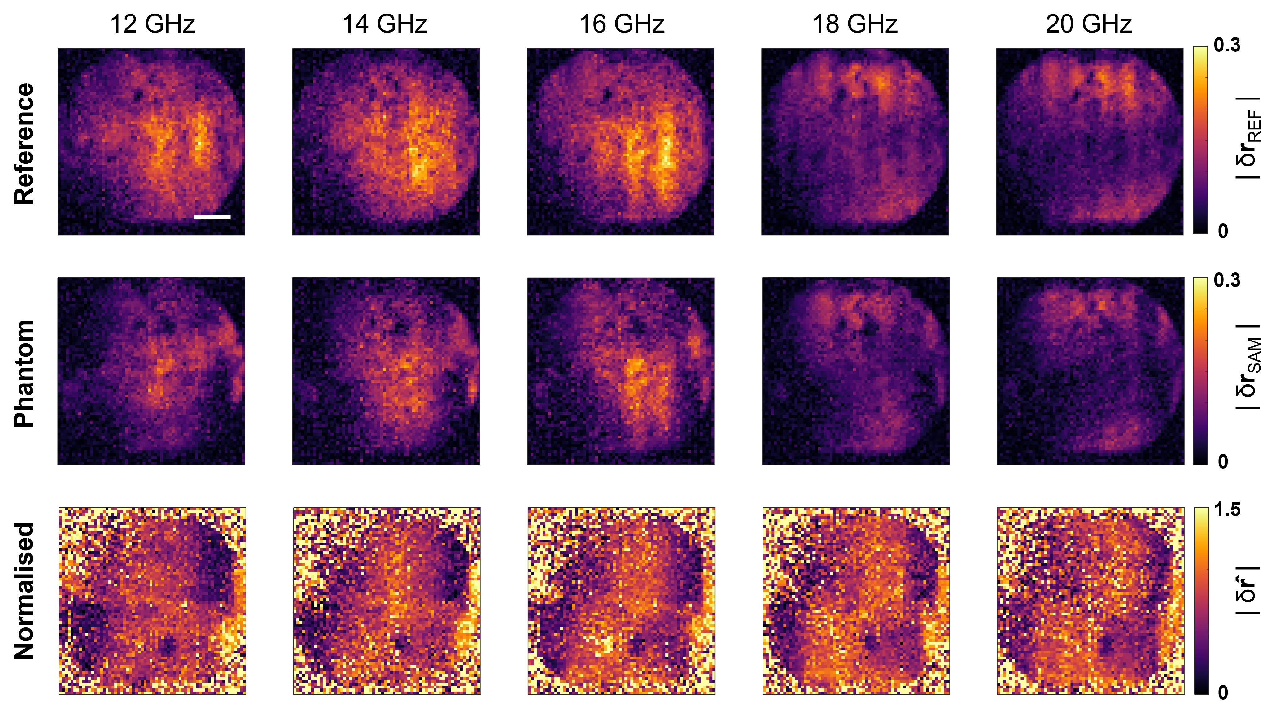
The device works like a microwave 'lamp': it illuminates the removed tissue with invisible radiation, and a single pixel captures the reflected signal. Under the tissue lies a silicon wafer that changes its transparency to microwaves when exposed to light. This allows scanning a 10 by 10 centimeter area with a step of just one millimeter, like running your finger over a surface and feeling every bump.
The authors tested the method on gelatin models of tumors with varying water content. The device found and measured even a thin layer of diseased tissue 2 millimeters thick — exactly the threshold surgeons consider dangerous. Numerical simulations showed the method is not thrown off by individual differences in patients' tissues.
Such a scanner fits into an operating room and requires no lengthy setup. If trials on real tissues confirm the results, repeat surgeries due to unclean margins could become rare.
Culinary gelatin and gelatin for tumor phantoms are almost the same thing: it mimics soft tissues well in terms of water content and microwave permeability.