X-ray imaging without interference, like a photo without glare express
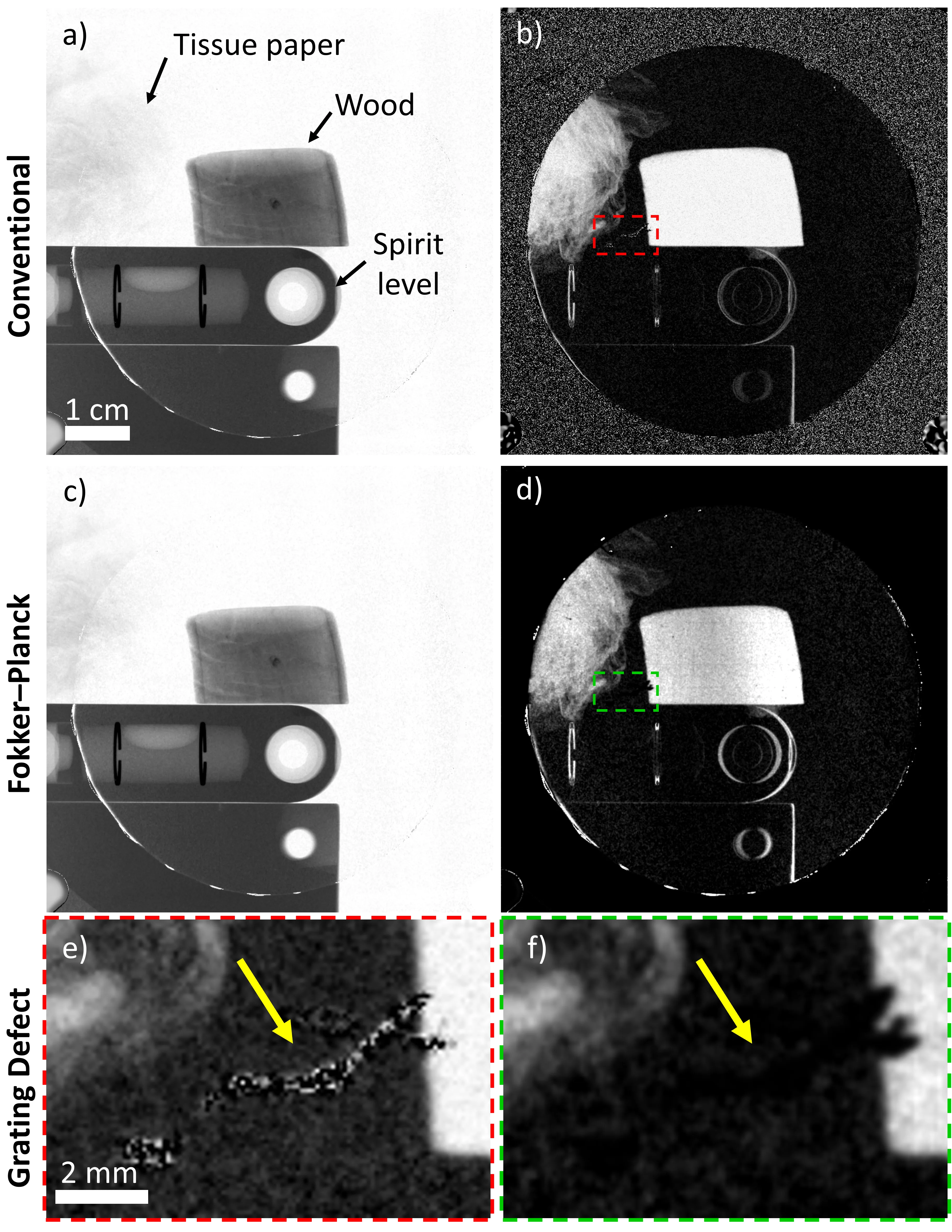
An X-ray machine with gratings takes not one but three pictures at once: a regular image, a map of beam deflections invisible to the eye, and an image of tiny structures that scatter light. Together, they help spot, for example, emphysema or pulmonary fibrosis. But there's a catch: to separate these three layers, you need a clever mathematical trick. For decades, the same one was used — fitting the data to a sine wave, as if laying a wave with the right rhythm over the image. The problem is that any scratch on the grating or a weak signal throws off that rhythm, and stripes appear on the image, like ripples from a stone thrown into water.
The authors of the new study took as their starting point an equation that describes how light, passing through matter, slightly changes direction and scatters — much like a flashlight beam in fog. This is the Fokker–Planck equation, and it can account for the random jolts each photon receives. Instead of fitting a sine wave, the algorithm looks at how smoothly brightness changes from pixel to pixel and figures out on its own where the real shadow is and where the junk from grating defects is. It's as if the image is wiped with a soft cloth, removing dust but leaving important details untouched.
This isn't just a cosmetic improvement. When an X-ray is taken quickly (for example, to avoid asking the patient to hold their breath for too long) the signal weakens, and noise can hide a tiny nodule. The Fokker–Planck-based algorithm extracts more useful information from such noise than the usual sine wave. It's like an experienced restorer who, from a few surviving brushstrokes, reconstructs the entire painting without inventing extra details.
The Fokker–Planck equation, which underlies the method, was originally devised to describe Brownian motion: those chaotic jumps of pollen in water that botanist Robert Brown once observed under a microscope.
In science fiction stories about future medical scanners, the idea of a device that sees through a person without any distortion often appears: this algorithm takes a step in exactly that direction.