How a Computer Learned to Measure Growing Organoids express
A biologist looks through a microscope at dozens of tiny cell spheres — spheroids. From these, miniature organ-like structures will later grow. The task is similar to a baker's job of checking whether the dough has risen evenly in each mold. Only here, instead of molds, there's a nutrient medium, and instead of dough, there are living cells. They cluster together on their own, and one experiment can yield a great many of these clusters.
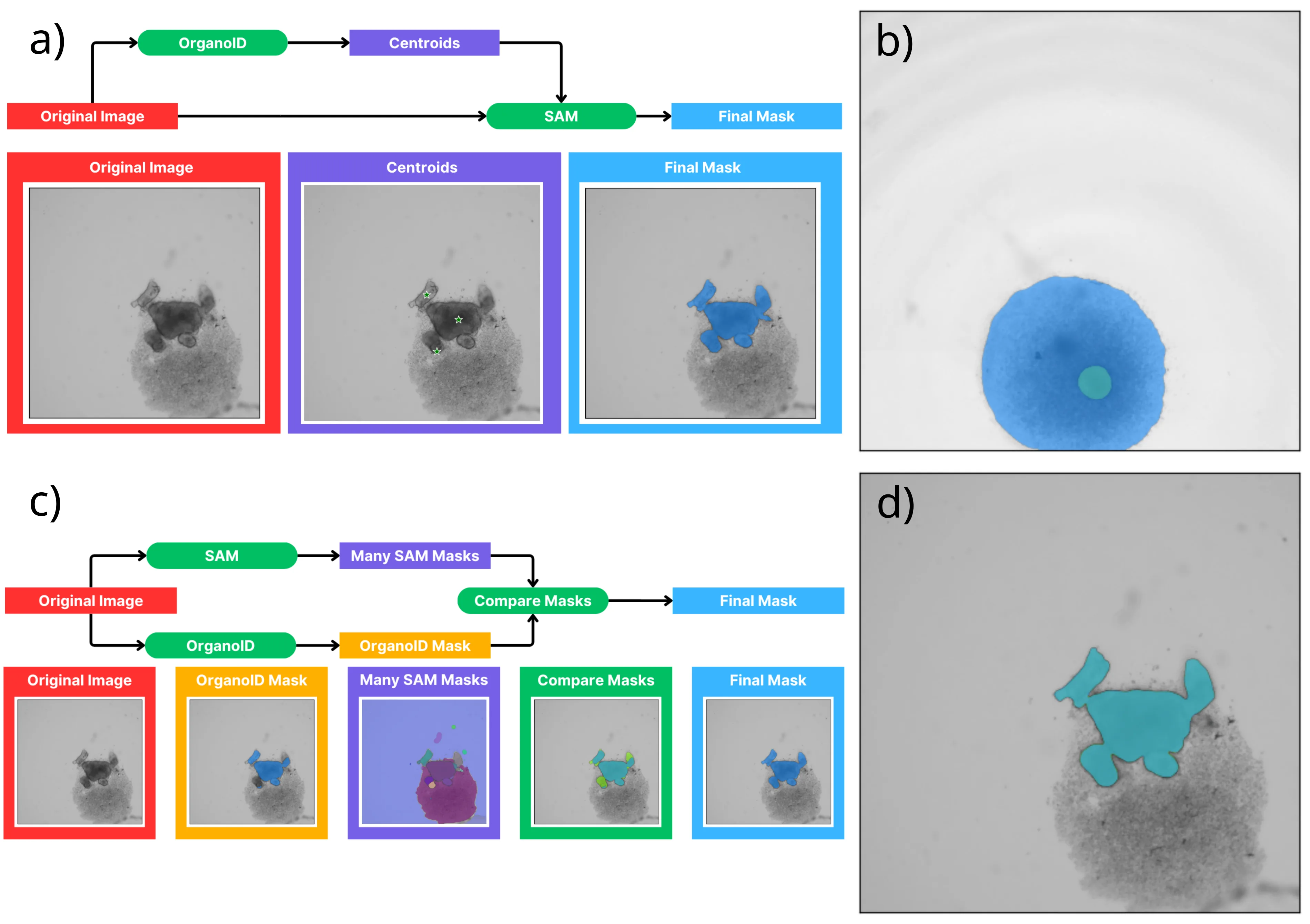
Previously, scientists manually outlined each sphere in images to measure its size and shape. This is time-consuming, tedious, and depends on who does the outlining: two people might draw the boundaries slightly differently. The authors of the new study decided to hand this routine over to a computer. They took the powerful universal model Segment Anything (SAM)—a sort of "Swiss Army knife" for segmenting objects in any image, and combined it with a tool specifically tailored for analyzing biological spheroids. The result is a hybrid that doesn't require lengthy retraining.
Tests showed that none of the existing tools, when used alone, performs consistently well across all images. But the new composite method delivers consistently accurate results for nearly all images — even the toughest ones where spheres clump together or lose clarity. Moreover, its accuracy is comparable to how two different people would outline the same sphere. Only the computer does it in seconds, not hours.
Now biologists have a fast and reliable "automatic measurer." It doesn't get tired or argue with colleagues about where to draw the line. Observing the development of mini-organs will become easier, and drug discovery and disease research a bit faster and more precise. Thus, the routine fiddling with contours gives way to live observation of how cells themselves build tiny organs.
Original abstract
as written by the authors arXiv:2605.03053
Organoids are complex, three dimensional, self-organizing cell cultures which manifest organ-like features and represent a powerful platform for studying human disease and developing treatment options. Organoid development is characterized by dynamic morphological and cellular organization, which mimic some aspects of organ development. To study these rapid changes over the course of organoid development, advanced imaging and analytical tools are critical to accurately monitor the trajectory of organoid growth and investigate disease processes. In this work, we focus on computer vision and machine learning techniques to automatically measure the size and shape of developing spheroids derived from pluripotent stem cells (iPSCs), which are typically the starting material for generating organoid cultures. To facilitate this task, we introduce a composite method that combines the Segment Anything Model (SAM), a general-purpose foundation model, with an existing domain-specific tool. This composite method is evaluated together with several existing tools by testing them on organoid image data and comparing with the results of manual image segmentation. We find that no single existing tool is able to segment the test images with sufficient accuracy across all test conditions, but the newly introduced composite method produces consistent and accurate results for all but a very small fraction of the most challenging images. Finally, we compare the accuracy of this method to the variability between manual segmentations by independent annotators (inter-observer variability) and find that by one measure it performs at the level of inter-observer variability and by others it performs very close to it.
Cell spheroids, from which organoids are grown, resemble an early embryo in shape and behavior at the very first stages of development, the cells also "negotiate" and self-organize without external commands.
In the movie "Blade Runner," artificial organs are created in test tubes — modern organoids can't yet replace a real liver, but they already help test drugs without animal experiments.