How Light Reveals the Fibers of Tissue express
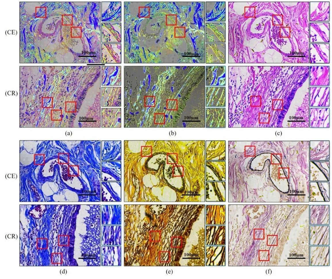
Inside our bodies, between cells, there is a support network — the extracellular matrix. It's like scaffolding made of proteins and carbohydrates that holds cells in place. When the fibers in this network change, it can be a sign of disease — for example, cancer or fibrosis.
Usually, to see these fibers, tissue is cut into thin slices and stained with special dyes. This is time-consuming and sometimes damages the sample. In the new work, scientists tried a different approach: they passed polarized light through the tissue — light whose waves oscillate in a single plane. Different fibers rotate the plane of light oscillation in different ways. By analyzing these changes, one can tell which fiber is in front of us.
To distinguish fiber types, the scientists used the UMAP method: it helps reveal structure in complex data, as if we were sorting similar objects into different piles. It turned out that collagen, elastic, and reticular fibers produce different polarization patterns. This means that by looking at the light, one can determine the fiber type without staining.
Such an approach could speed up diagnosis: instead of hours of staining, a quick analysis under a polarized microscope. In the future, this will complement traditional methods and help doctors more accurately assess tissue condition.
Polarized light is used not only in microscopy: for example, bees navigate by the polarization of the sky even on cloudy days.