Separating Brain Scans: Where Disease Ends and Distortion Begins express
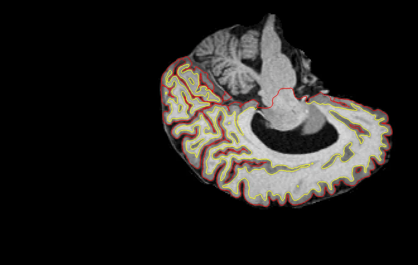
Two scans of the same brain taken at different times are like photographs of the same house before and after renovation. To see what changed, you overlay them and look at the differences. But an MRI scanner is not a perfect camera: it adds its own distortions, as if the photo were taken through slightly warped glass. Previously, programs could not tell which changes were real and which were from the machine.
The neural network DisMorph learns from fully synthetic examples where it is known in advance which part of the distortion is technical and which is anatomical. It then decomposes the differences between two scans into two streams: one accounts for scanner artifacts, the other for real tissue changes. This approach is reminiscent of a restorer who separates cracks from old paint to understand what was originally in the painting.
On pairs of scans from patients with Alzheimer's disease, the method detected changes in brain structures associated with the disease while also pointing to residual distortions after standard correction. It is as if the doctor received not one blurry image but two clear ones: one showing the brain itself, the other showing the flaws in the imaging.
This approach paves the way for more accurate measurements of brain changes in clinics where machines and settings are not always uniform. Instead of guessing whether what appears on the scan is disease or artifact, the doctor will be able to see them separately.
Original abstract
as written by the authors arXiv:2608.08173v1
Longitudinal MRI enables sensitive measurement of structural brain change for studying aging and neurodegenerative disease. Deformable image registration is a key tool for estimating such change by computing a dense deformation that captures geometric differences between longitudinal scans. However, MRI scanners introduce geometric distortions that vary across acquisition systems and protocols, such as gradient non-linearity (GNL) distortion. Existing registration methods estimate a single field that conflates biological and technical effects, potentially biasing downstream morphometric measurements if distortions remain (partially) uncorrected. We propose $$\texttt{DisMorph}$$, a registration framework trained entirely on synthetic data that explicitly decomposes longitudinal deformation into technical and anatomical transforms. It predicts two dense deformations, each encoding one effect. During training, a novel generative model synthesizes both effects separately to provide disentanglement supervision, while domain randomization promotes generalization across imaging protocols. We evaluate our method in three complementary settings. On simulated data with known ground truth, our method detects anatomical change more accurately and consistently than conventional registration. On real image pairs that differ only by GNL distortion, our method assigns most geometric change to the distortion field, demonstrating specificity in the absence of anatomical change. On longitudinal Alzheimer's disease (AD) pairs, our method detects anatomical change in AD-related brain structures while identifying residual distortion left after standard correction. By disentangling MRI-induced distortion from biological change in the longitudinal deformation, our method paves the way for more accurate longitudinal morphometry in clinical settings where maintaining acquisition consistency is challenging.
Gradient nonlinearity is a distortion in which straight lines in an image appear curved, like a reflection in a funhouse mirror. It arises from imperfections in the scanner's magnetic field.
In the series "Black Mirror," there is an episode where memories are recorded and replayed, but the technology distorts details. DisMorph tackles a similar problem: separating technical artifacts from real changes, only for brain scans.