Light from Within: How to See 12 Centimeters Deep into the Body express
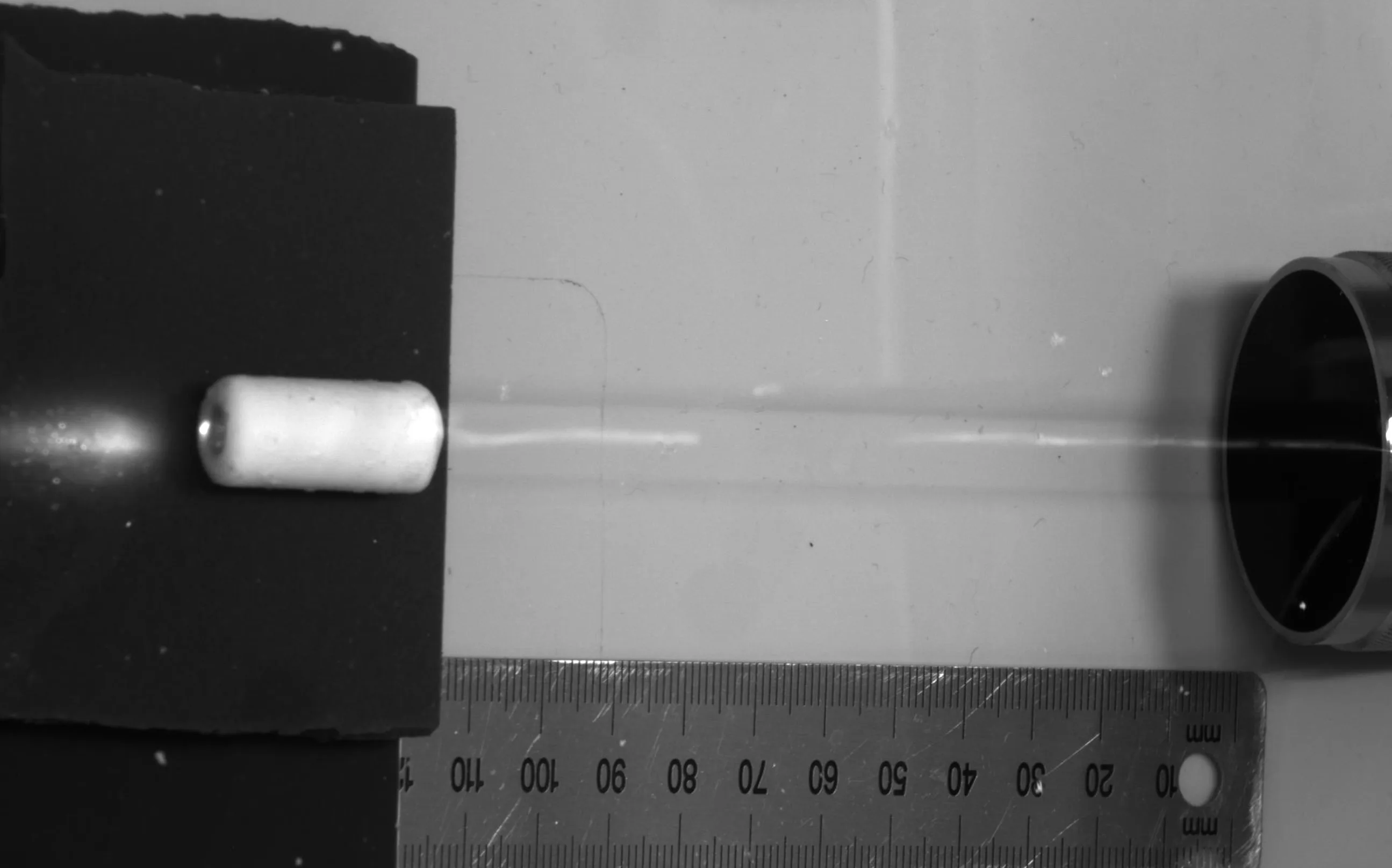
Light is a great tool for studying tissues, but it fades fast deep inside the body. Within a few millimeters, photons scatter so much that you can't make out anything. Scientists came up with a workaround: instead of shining light from outside, deliver a tiny light source right inside. The challenge is how to power that source without wires.
The solution came from ultrasound. Low-frequency sound waves pass through the body with almost no loss: that's why ultrasound imaging can see deep organs. In the new device, ultrasound not only "lights" the way but also carries energy: a compact receiver converts acoustic vibrations into electricity, which powers a pulsed laser diode. The diode emits short flashes of light, tissues absorb them, heat up by fractions of a degree, and expand — generating a weak ultrasound wave. A sensor outside picks up that wave, and from these signals, a map of optical absorption is reconstructed.
The researchers tested the method on a phantom 12 centimeters thick — about the depth of an adult's abdominal cavity. The device, the size of a pill, handled the job, even though ordinary external light wouldn't have made it a tenth of that distance. In the future, this approach could let us look into the gastrointestinal tract, blood vessels, and other areas that neither an endoscope nor external lighting can reach.
Original abstract
as written by the authors arXiv:2601.01667
Photoacoustic tomography leverages ultrasound's deep tissue penetration to retrieve optical absorption contrast well beyond the optical diffusion limit. Conventional photoacoustic systems rely on externally delivered light and are therefore constrained by optical attenuation, limiting imaging depths to several centimeters. Here, we overcome this constraint using a compact, acoustically powered device that provides optical excitation directly from within the target medium. By exploiting the weak attenuation of low-MHz ultrasound, acoustic energy is transmitted through tissue to wirelessly power a pulsed laser diode. The emitted light pulses generate photoacoustic signals that encode local optical absorption at clinically relevant depths, which could enable imaging in regions such as the gastrointestinal tract that are inaccessible to surface-based illumination. We demonstrate this approach by imaging through a 12 cm thick phantom, establishing a pathway toward deep-tissue photoacoustic imaging.
Ultrasound at around 1 MHz attenuates in soft tissues hundreds of times slower than visible light, so it's used both for diagnosis and for delivering energy.
The idea of "swallowing a light source" recalls capsules from science fiction films that travel through the body and transmit images to a doctor's screen.