Proteins bend the membrane the way a spoon bends the film on milk express
The membrane of a living cell is not a rigid shell but a pliable film with thousands of proteins embedded in it, like raisins in dough. When a protein changes shape or the cell moves, the film bends: sometimes barely noticeable, like the surface of water when a speck of dust falls on it, and sometimes strongly, like a sheet when you stretch your legs under it. Until now, calculations described only weak bends, but in a real cell, bends can be deep: during division, particle uptake, or formation of transport vesicles. The authors of the new work have for the first time tackled strong bends and found something unexpected.
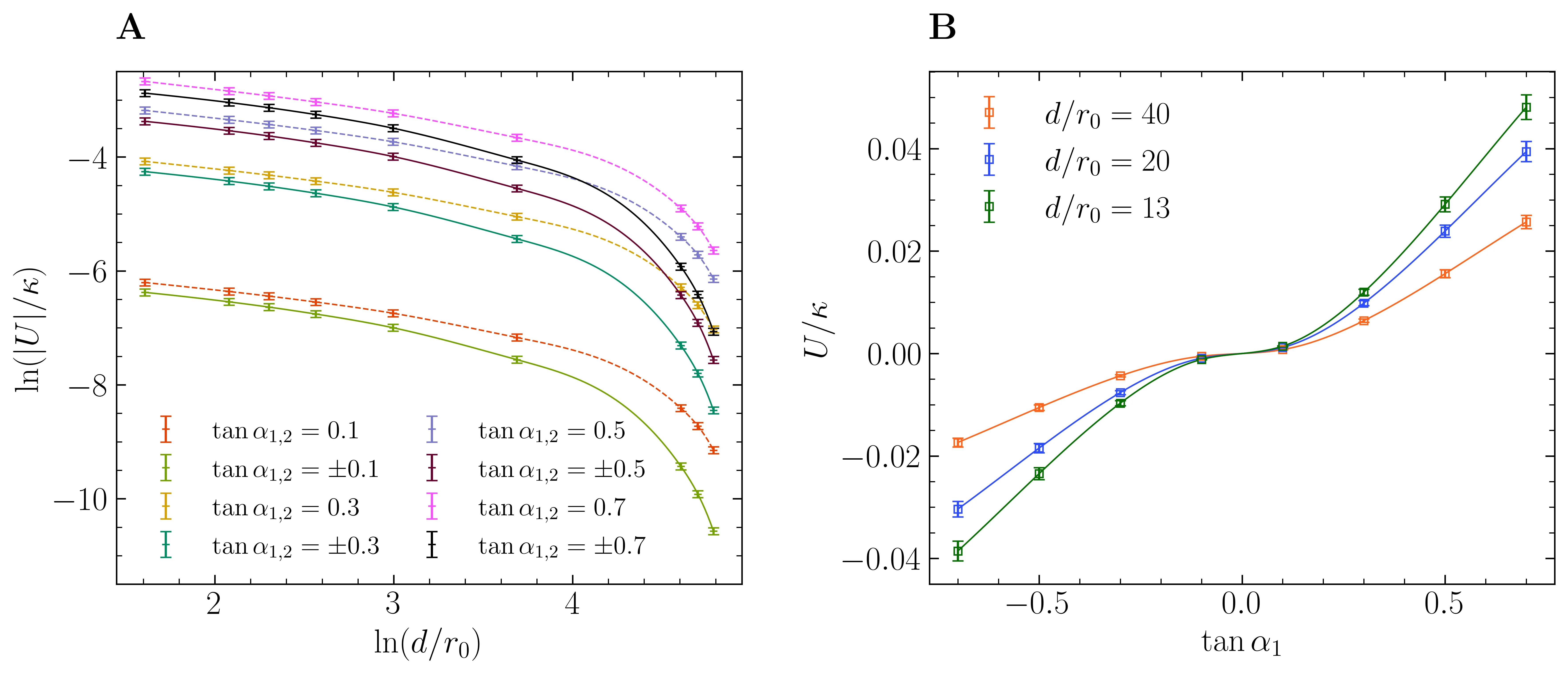
They used a computer model where the membrane and proteins are divided into many small elements, like a mosaic. It turned out that the force with which the bent membrane pulls a protein up or down is not monotonic: it first increases, then decreases. This is like stretching a rubber band with a bead glued to it — the farther you pull the bead, the stronger the band resists, but at some point the band slips out from under your fingers, and the force drops. Moreover, this pattern does not depend on the protein's shape — whether it's a cone or a cylinder. For two proteins next to each other, the calculation showed: if both are tilted in the same direction, they repel; if in opposite directions, they attract. This resembles the behavior of electric charges, except here the 'charge' is the protein's tilt.
Such calculations help understand how proteins cluster on the cell surface, how the cell sorts cargo and sends it to the right compartments. All of this is the basis of life, but until now we have only seen weak shadows of these processes. Now that strong bends are calculated, the picture becomes three-dimensional.
The force with which the membrane pulls on a protein can first increase and then decrease, as if the film 'gets tired' of holding the protein under strong bending.