Full-Field Fluorescence Tomography: Chemistry in 3D Without Scanning ✛
Context
X-ray fluorescence tomography typically requires point-by-point scanning with a focused beam, which is slow and limits co-registration with full-field structural tomography. Around this work in the archive are methods for improving X-ray imaging: dose reduction, sharper resolution, quantum microscopes. This paper stands apart: it is the first to demonstrate an experimentally calibrated three-dimensional full-field fluorescence tomography compatible with phase contrast.
Methods
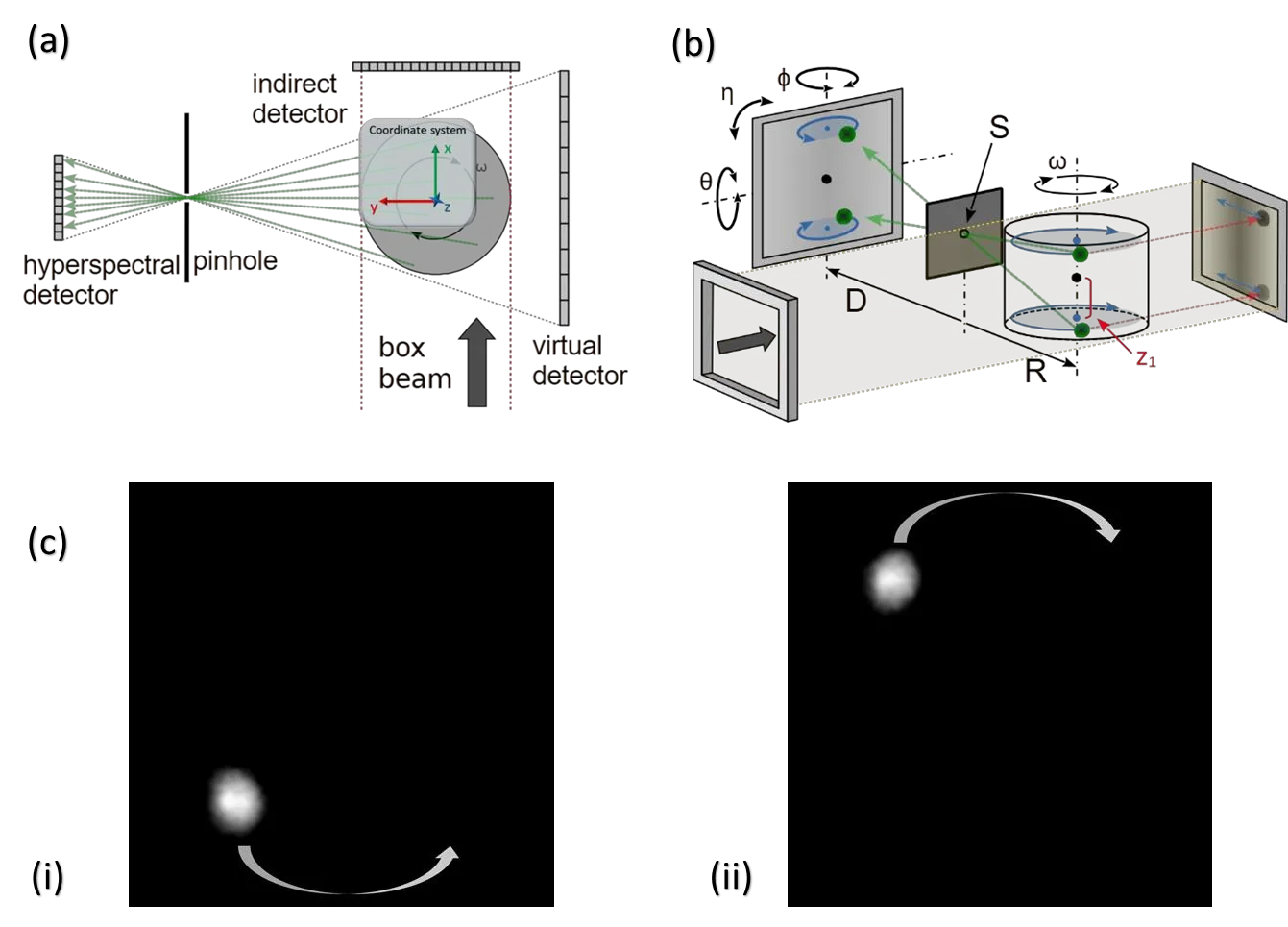
The experiment was conducted at the ESRF synchrotron, beamline ID19, with a polychromatic beam of mean energy 59.25 keV. The sample is illuminated entirely, and the fluorescent emission passes through a 200 μm diameter pinhole and is recorded by the HEXITEC pixel detector with 250 μm pitch. The geometry is equivalent to a virtual cone beam, where the pinhole acts as a point source. Calibration is two-step: first, geometric parameters are estimated from projections of a calibration sphere, then refined by minimizing reconstruction error. Phase-contrast tomography was acquired with an effective pixel size of 3.1 μm and 5000 projections.
Results
F3CT reconstructed three-dimensional elemental distributions without raster scanning. For the silver-stained zebrafish embryo, a volume with Ag Kα signal (22.16 keV) was reconstructed, showing uneven distribution of the stain in soft tissues. For the geological core, Fe-sensitive and Ba-sensitive volumes were separated: the Ba signal is localized in the granite, while the Fe signal is distributed in the serpentinite. The spatial resolution of the fluorescence is about 150 μm, lower than phase contrast but sufficient for chemical mapping.
Implications
This work differs from neighboring methods in that it is the first to combine full-field illumination, a hyperspectral detector, and calibrated virtual cone-beam reconstruction for three-dimensional fluorescence tomography. This opens the way to correlative 'structure + chemistry' imaging in a single experiment without changing geometry.
Future development
Prospects involve replacing the single pinhole with coded apertures to increase throughput and resolution, as well as a new generation of HEXITEC detectors with megacount-per-second rates. This will enable in-situ and operando studies of dynamic processes, such as elemental migration in batteries.
Impact
The method is applicable in biology, geology, materials science, and cultural heritage, wherever non-destructive volumetric chemical mapping is needed. It also lays the groundwork for future low-dose imaging of biological tissues.
Next steps
Development of coded apertures and deconvolution algorithms to improve resolution and sensitivity. Integration with fast detectors for time-series studies.
Key open problems
In X-ray imaging, the challenge remains to obtain both high spatial resolution and chemical sensitivity without excessive dose. F3CT addresses this through parallel detection, but it is limited by the pinhole resolution and the detector's sensitivity to low energies.
The knowledge machine's view
This section is addressed to the author of the paper and their colleagues. Neighbouring works were found by meaning-based search across our archive, not by word overlap. We do not review: we show the field around the work.
What the work does
This paper presents full-field fluorescence computed tomography (F3CT) at the ESRF synchrotron: the sample is illuminated as a whole, the fluorescence signal passes through a pinhole and is recorded by the HEXITEC hyperspectral detector, and the volume is reconstructed using a virtual cone-beam model with a two-step calibration. The method was validated on a zebrafish embryo and a geological core, yielding three-dimensional distributions of silver, iron, and barium; in the same geometry, phase-contrast tomography was also acquired for structural context.
Where its strength is
The main strength is the combination of full-field illumination, an energy-sensitive detector, and a calibrated virtual cone-beam reconstruction, which allows chemical mapping of the volume without raster scanning and in the same geometry as phase contrast.
Where it could go next
- It might be interesting to see how dose-reduction and data-acquisition acceleration methods described in a neighboring work on full-field imaging without line-by-line scanning could be adapted for F3CT to broaden its applicability to radiation-sensitive samples.based on: X-Ray Vision Through a Sample Without Point-by-Point Scanning arXiv:2604.10671
- There is a nearby work on improving resolution using quantum methods; perhaps some of those ideas could be applied to fluorescence photon detection to enhance F3CT spatial resolution without increasing dose.based on: Quantum trick boosts microscope resolution by 5 times arXiv:2605.05961
- The archive contains a work on correlative imaging using annihilation photons; combining F3CT with such methods might provide additional contrast for biological tissues.based on: The Quantum Waltz of Annihilation: What Photons Reveal About Tissues arXiv:2607.12182
Worth trying
- Test iterative reconstruction with regularization that uses phase-contrast data as prior information to improve the fluorescence volume.
- Investigate how reducing the pinhole size and increasing the number of projections affects resolution and experiment time.
- Consider using a multimodal detector that records both fluorescence and scattering simultaneously to obtain additional information about the sample matrix.
Why this may matter
Such correlative imaging of structure plus chemistry in a single experiment could be useful for studying biomineralization, drug distribution in tissues, and geochemical processes where spatial correlation of elemental composition with microstructure matters.
Nearby works in our archive
- X-Ray Vision Through a Sample Without Point-by-Point Scanningsimilarity 0.57
- A Medical 3D Scanner That Learns to See It All at Oncesimilarity 0.52
- Live Cells at Video Speedsimilarity 0.51
- A Hologram from a Speckle of Lightsimilarity 0.50
- Light Pierces Milk: Lensless Microscope Sees Through Murky Mediasimilarity 0.50
- Quantum trick boosts microscope resolution by 5 timessimilarity 0.50
- The Quantum Waltz of Annihilation: What Photons Reveal About Tissuessimilarity 0.50
The archive is sparse around this work: the closest review is only 0.57, against a usual 0.64. It looks like few have drilled nearby.
The accumulation time for one fluorescence projection was 150–170 seconds, with 121 projections in total, about 5.5 hours per dataset. The phase-contrast tomography with 5000 projections took a comparable time.
The idea of seeing the chemical composition of a volume without destroying it evokes the tricorder from 'Star Trek', which scans an object and gives an elemental analysis. F3CT is a step toward such a device, though it still requires a synchrotron.
Key numbers
- effective fluorescence pixel: 144 μm
- fluorescence spatial resolution: ~150 μm
- accumulation time per projection: 150–170 s
- number of angular projections: 121
- beam energy: 59.25 keV