Diagnosis by Reaction: What Healthy Brain Tissue Reveals About Disease ✛
Context
This work stands apart: the nearest neighbors in the archive focus on medical image segmentation, brain cancer detection, and weakly supervised learning, but none have tackled diagnosis from reactive tissue without obvious lesions. Existing methods can classify tumors by histology, but they all assume tissue with clear pathology. Here the task is fundamentally different: the signal is weak, diffuse, and hidden in tissue that pathologists traditionally regard as non-diagnostic.
Methods
From the University of Münster archive, the authors took 245 hematoxylin-and-eosin-stained sections from 186 patients with confirmed diagnoses. To isolate reactive tissue, they manually removed tumor regions or used biopsies deemed non-diagnostic but with a subsequent confirmed diagnosis for the same patient. Each section was tiled into patches, encoded with four different "foundation" histology models (UNI2-h, Virchow2, Prov-GigaPath, H-optimus-0), and then patches were aggregated via an attention mechanism into a single section vector. Training was done at three levels: tumor/inflammation/control, glioma/metastasis, and glioma subtype or metastasis origin.
Results
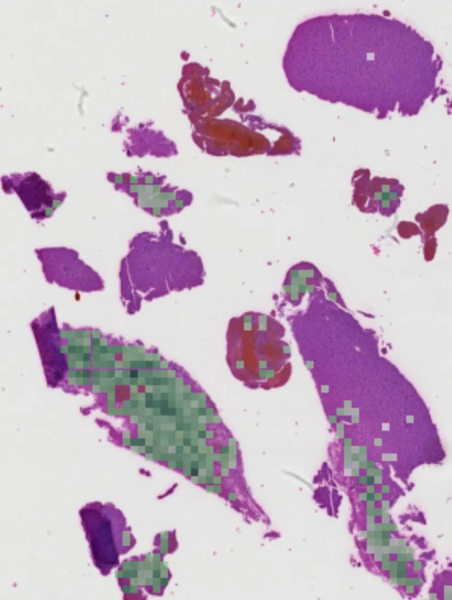
The main surprise: the coarse classification of tumor/inflammation/control is almost entirely explained by section size. Logistic regression on just the logarithm of patch count achieved a balanced accuracy of 0.782, compared to 0.798 for the best model. But on finer tasks (glioma subtype and metastasis origin), size no longer helped: the baseline dropped to chance level, yet models still classified significantly better (p≤10⁻⁴ in permutation tests). Meanwhile, none of the four encoder models differed statistically from one another: all pairwise differences vanished after correction for multiple comparisons. Model attention was partly drawn to blood introduced during tissue collection: for Virchow2, such patches made up 27% of the top-5, while for UNI2-h only 4%.
Implications
This work fits within weakly supervised histopathology but differs in its object: instead of obvious lesions, it targets reactive tissue that is usually discarded as uninformative. It is the first test of whether disease signals persist in "empty" parenchyma and can be extracted automatically. At the same time, the paper introduces a mandatory control, a baseline based solely on section size, which exposes false success on the coarse task. For the community, this is a signal: computational pathology benchmarks must check whether models exploit sample preparation artifacts.
Future development
The topic could evolve toward finer representations working at the level of individual nuclei rather than 256×256-pixel patches. It would also be promising to combine weakly supervised histology with spatial transcriptomics of the same reactive regions, linking morphological features to specific cellular programs. That would turn a purely predictive model into a tool for understanding the biology of gliosis.
Impact
The results will affect model evaluation protocols in computational pathology by adding a control for sample preparation artifacts. In the long run, the method could help neuropathologists extract diagnostic information from biopsies currently considered unsuccessful.
Next steps
Reproducibility must be tested on multicenter data with different scanners and staining protocols. Then, an analysis with a fixed patch budget and stratification by section origin is needed to separate morphology from technical artifacts.
Key open problems
The paper directly addresses the unresolved problem of diagnostic uncertainty in neuropathology: up to 14% of brain biopsies contain no tumor tissue, forcing clinicians to tell patients that the cause of their disease is unknown. This work shows that even such samples carry hidden information, but extracting it requires new approaches to image analysis and artifact control.
The knowledge machine's view
This section is addressed to the author of the paper and their colleagues. Neighbouring works were found by meaning-based search across our archive, not by word overlap. We do not review: we show the field around the work.
What the work does
The study tested whether a diagnostic signal can be extracted from reactive brain tissue that contains no tumor cells. Weakly supervised models with different encoders, attention-based patch aggregation, and control for the slide-size artifact were trained on 245 histological sections. After removing this artifact, the models distinguished subtle pathologies significantly better than chance, but the choice of encoder did not affect the results.
Where its strength is
A strong point is the clear protocol with a mandatory baseline on slide size, which exposes spurious success on coarse classification and confirms the reality of the signal on fine-grained tasks. This sets a standard for checking tissue preparation artifacts in computational pathology.
Where it could go next
- It might be interesting to see how attention interpretation methods from the work on cells that 'communicate with light' could help identify informative patterns in reactive tissue and understand which structures carry the signal.based on: Cells Communicate with Light arXiv:2510.05792
- A related approach uses graph neural networks for melanoma detection: a graph representation of patches could capture spatial relationships in tissue that are lost with attention-based aggregation.based on: How Graphs Find Melanoma on Your Skin arXiv:2604.03710
- The work on predicting chemotherapy response from MRI used multimodal data; adding radiomics or clinical features might strengthen the weak histological signal in your task.based on: How MRI Predicts Chemotherapy Response in Glioblastoma arXiv:2601.07035
Worth trying
- Test whether the signal is robust to patch sizes or overlapping patches.
- Use a graph neural network on patches to account for the spatial organization of reactive tissue.
- Add clinical variables (age, location) as additional features to the slide-level vector.
Why this may matter
If the signal truly persists in reactive tissue, it opens the door to diagnosis from biopsies currently considered non-informative and could increase the diagnostic value of each procedure.
Nearby works in our archive
- Cells Communicate with Lightsimilarity 0.54
- How Graphs Find Melanoma on Your Skinsimilarity 0.52
- Inside the Cell: How Cancer Dodges the Drugsimilarity 0.52
- Tongue Cancer: How a Scan Reveals the Tumor's Personalitysimilarity 0.52
- How MRI Predicts Chemotherapy Response in Glioblastomasimilarity 0.51
- When a Test's Silence Speaks Louder Than Numberssimilarity 0.51
The archive is sparse around this work: the closest review is only 0.54, against a usual 0.64. It looks like few have drilled nearby.
Models with identical accuracy can focus on completely different regions of a slide: one fixates on hemorrhages from tissue collection, another on subtle glial changes. In other words, a "correct" answer does not guarantee that the model found a biologically meaningful feature.
The idea of reading disease from the body's reaction rather than the pathogen itself echoes stories of diagnosis by indirect clues, like in the TV series "House M.D.", where the team diagnoses based on subtle symptoms rather than an obvious cause.
Key numbers
- up to 14% of brain biopsies contain no tumor tissue: rate of non-diagnostic biopsies
- 245 sections from 186 patients: dataset size
- 27% vs 4%: share of blood patches among top-5 attention for Virchow2 and UNI2-h
- p≤10⁻⁴: significance of difference from chance for all tasks
- 0.782 vs 0.798: accuracy of size baseline and best model on the coarse task