The Living Lens: How MRI Sees Muscles in Motion express
A photographer snaps a runner: to avoid a blurry shot, a short shutter speed is needed. An MRI scanner is like that photographer, but its 'shutter speed' is long — by the time it captures, the muscle has already changed. That's why dynamic MRI (a series of volumetric frames in motion) is a tough challenge: data are scarce, and motion doesn't wait.
A team of researchers has prepared an open dataset: nine healthy volunteers, with a pneumatic cuff on the thigh that compresses muscles in a controlled, repeatable way. For one type of deformation, they recorded both incomplete (fast) data and full reference images. The other three deformations were recorded only as incomplete data, as in a real clinical setting.
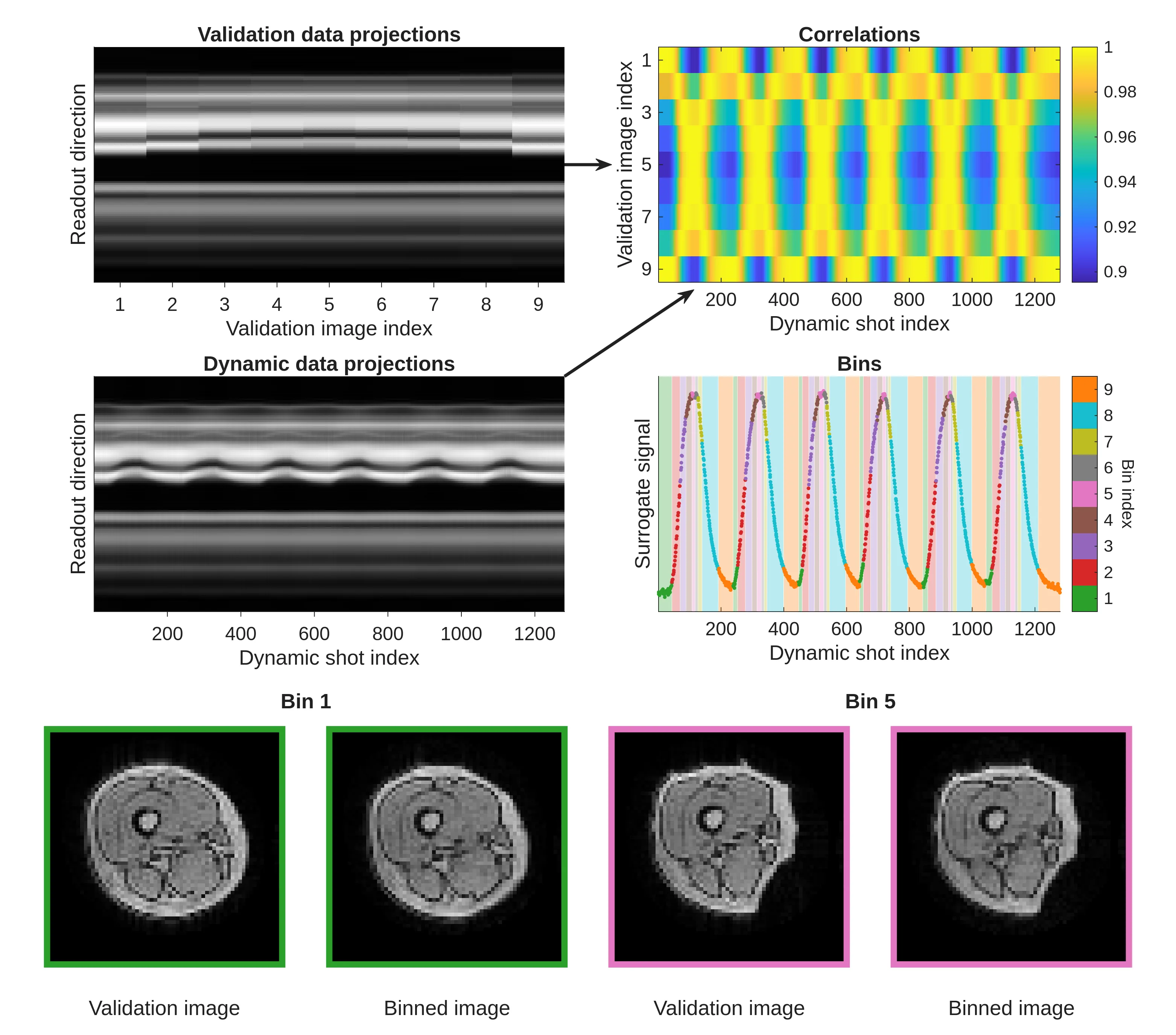
To demonstrate how to use the dataset, the authors applied a simple method of sorting data by motion phases and reconstructed the images. The result matched the reference, so the dataset is suitable for training and testing more advanced algorithms.
Now any researcher can download this data and test their own dynamic MRI reconstruction method. It's a step toward turning the scanner into a video camera for internal organs, so doctors see not a frozen image, but living motion.
Original abstract
as written by the authors arXiv:2602.08918
Dynamic volumetric MRI provides valuable information on in vivo motion and biomechanics, with applications spanning cardiac, musculoskeletal, or pulmonary imaging, amongst others. Developing reconstruction methods for time-resolved volumetric MRI is challenging due to the inherently slow acquisition process of MRI, which makes it an active area of research. However, in vivo validation of these methods remains challenging due to the lack of publicly available datasets with fully sampled ground-truth images. Here, we present a publicly available in vivo dataset designed to facilitate the development and validation of dynamic volumetric MRI reconstruction algorithms. Controlled and repeatable deformations of the muscles in the thigh were induced using a pneumatic pressure cuff, enabling the acquisition of both undersampled dynamic data and fully sampled validation images. The dataset comprises multichannel undersampled k-space data from nine healthy volunteers across four different dynamic deformations, with fully sampled validation data for one deformation. Additionally, an anatomical reference scan and muscle segmentation masks are provided for each subject. To illustrate a possible image reconstruction and validation approach, a binning-based reconstruction was performed on the undersampled data from six dynamic repetitions. The resulting images were consistent with the corresponding fully sampled validation images. This dataset offers possibilities for validating and advancing time-resolved volumetric MRI reconstruction methods.
An MRI scanner measures the signal not in image space, but in so-called k-space, like the notes of a musical score, from which the picture-melody is later reconstructed.