The Living Lens: How MRI Sees Muscles in Motion express
A photographer snaps a runner: to avoid a blurry shot, a short shutter speed is needed. An MRI scanner is like that photographer, but its 'shutter speed' is long — by the time it captures, the muscle has already changed. That's why dynamic MRI (a series of volumetric frames in motion) is a tough challenge: data are scarce, and motion doesn't wait.
A team of researchers has prepared an open dataset: nine healthy volunteers, with a pneumatic cuff on the thigh that compresses muscles in a controlled, repeatable way. For one type of deformation, they recorded both incomplete (fast) data and full reference images. The other three deformations were recorded only as incomplete data, as in a real clinical setting.
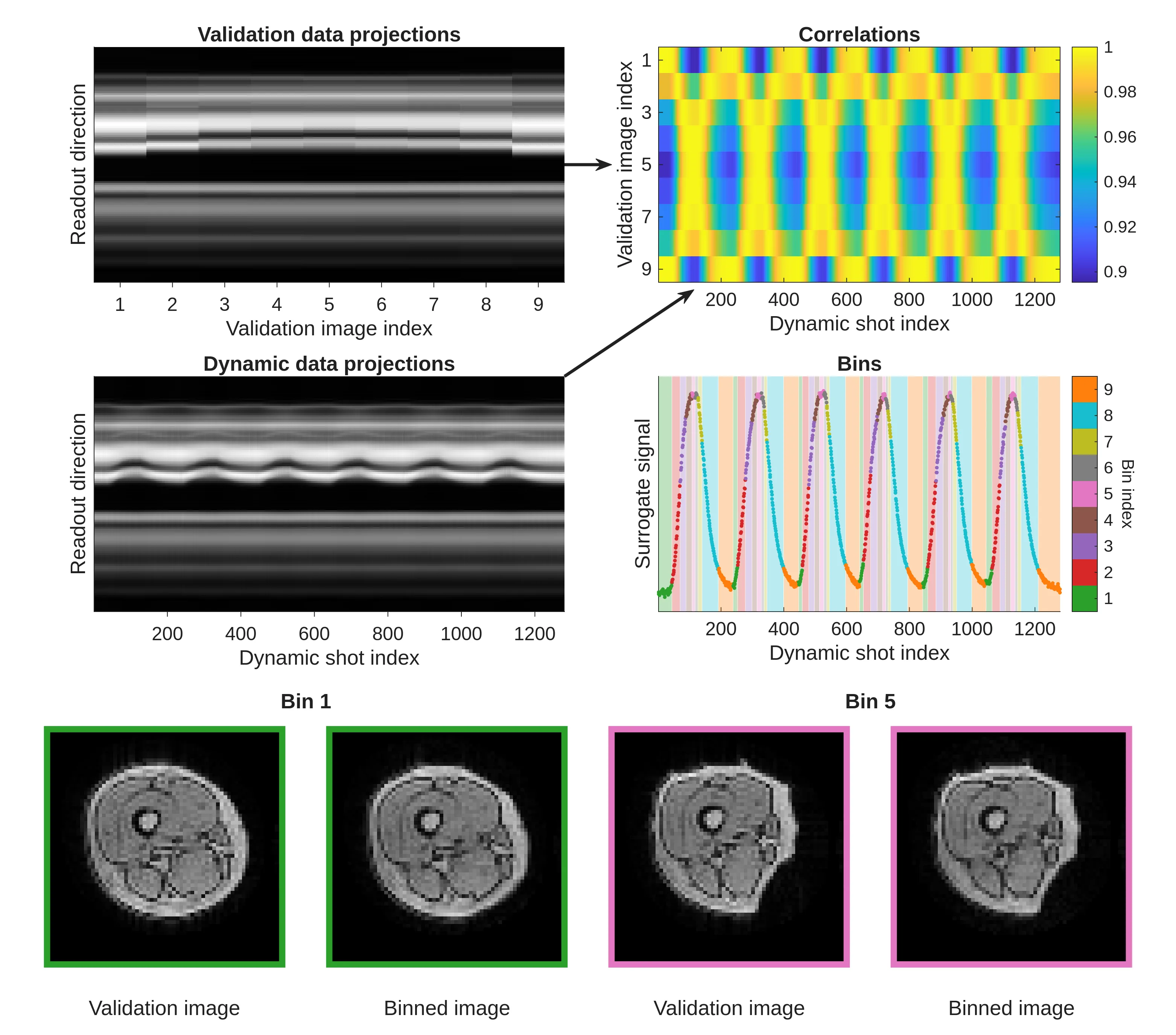
To demonstrate how to use the dataset, the authors applied a simple method of sorting data by motion phases and reconstructed the images. The result matched the reference, so the dataset is suitable for training and testing more advanced algorithms.
Now any researcher can download this data and test their own dynamic MRI reconstruction method. It's a step toward turning the scanner into a video camera for internal organs, so doctors see not a frozen image, but living motion.
An MRI scanner measures the signal not in image space, but in so-called k-space, like the notes of a musical score, from which the picture-melody is later reconstructed.