Seeing the Invisible: A Clear View of Transparent Cells express
A glass marble in a glass of water is nearly invisible, but if you look through it at a checkered tablecloth, the lines behind it appear distorted. Transparent cells behave the same way: light passes through them but is slightly slowed and changes direction. A regular microscope can't catch such subtle changes: you need dyes, which often kill living cells.
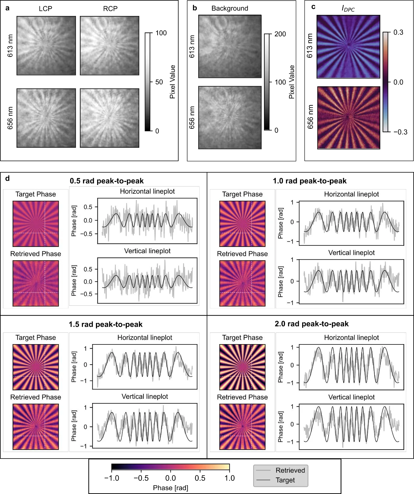
Engineers made a plate thinner than a human hair and covered it with a pattern of tiny pillars. When light passes through a cell and this plate, the pillars act like millions of tiny lenses: they deflect the rays exactly as much as the cell slowed them. One click of the shutter, and you get two images showing how light bent up-down and left-right. From these two pictures, a computer assembles a precise map: where the cell is thicker, where denser, where its nucleus is.
The method was tested on HeLa cells — the famous cell line that has been living in labs since 1951. Without a single drop of dye, the internal structure of the cells is visible in the images, and the measured thickness matched data from other studies. Such plates can be placed in regular microscopes or even in portable devices, so doctors and biologists can see living cells right at the patient's bedside.
The HeLa cells used in this work are descendants of Henrietta Lacks's tumor cells. They divide so actively that over decades their total mass has exceeded Henrietta's own mass by millions of times.
In the movie Gattaca, characters get a diagnosis from a drop of blood, such fast and accurate diagnostics without damaging cells becomes closer with compact phase microscopes.