Rethinking Light Distribution Reveals the Invisible express
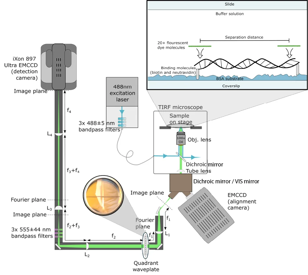
A conventional light microscope hits a fundamental limit: it cannot distinguish two points closer than 200 nanometers — roughly the thickness of a soap bubble divided by a hundred. To see deeper, scientists have made molecules in the sample blink or fade, but these tricks are slow and harmful to living cells. The new device, called the Structured Detection Microscope, takes a different approach.
It doesn't touch the sample itself; instead, it alters the shape of the light spot captured by the camera. Light from neighboring molecules that would normally merge into one blurry spot is spread across different parts of the sensor. It's like recording two voices in a noisy room not with a single microphone but with two placed in opposite corners: each picks up its own share. As a result, the device can distinguish labels on DNA rulers spaced 50 nanometers apart and achieves clarity down to 40 nanometers — five times better than the limit.
This approach paves the way for observing living cells without phototoxicity — the light-induced damage that degrades cells under a microscope. Now biologists can track proteins and DNA in real time without fearing that a bright beam will kill the cell.
Original abstract
as written by the authors arXiv:2604.00413
Super-resolution microscopy is crucial for imaging sub-wavelength biological structures. However, most techniques rely on nonlinear saturation or stochastic switching of emitters, limiting imaging speed and increasing phototoxicity. Here, we achieve deep super-resolution without employing saturation or stochastic dynamics, instead using a form of spatial mode demultiplexing. By shaping the point-spread function of the emitted light, our Structured Detection Microscope (SDM) redistributes information away from high shot-noise regions of the image, enhancing sensitivity to sub-diffraction emitter separations in two-dimensions and without mode-sorting optics. Implementing SDM within a high-numerical aperture total internal reflection fluorescence microscope, we demonstrate imaging of fluorophores attached to DNA nanorulers with separations as small as 50 nm at resolutions surpassing 40 nm - fivefold below the diffraction limit. This shows that spatial mode demultiplexing can achieve far sub-wavelength resolution and is applicable to biologically relevant samples. By enabling super-resolution biomolecular imaging without emitter saturation and stochasticity, our work opens the door to better understanding biological structure, function and dynamics.
The optical resolution limit that the authors overcame was described back in 1873 by Ernst Abbe. It was considered insurmountable for nearly a century and a half.
In the series 'The Expanse,' ultra-precise sensors allow ships to see through asteroids. The new microscope is a step toward seeing through cell membranes without destruction.