Diamond Sensor Catches Magnetic Signal from a Living Cell ✎ express
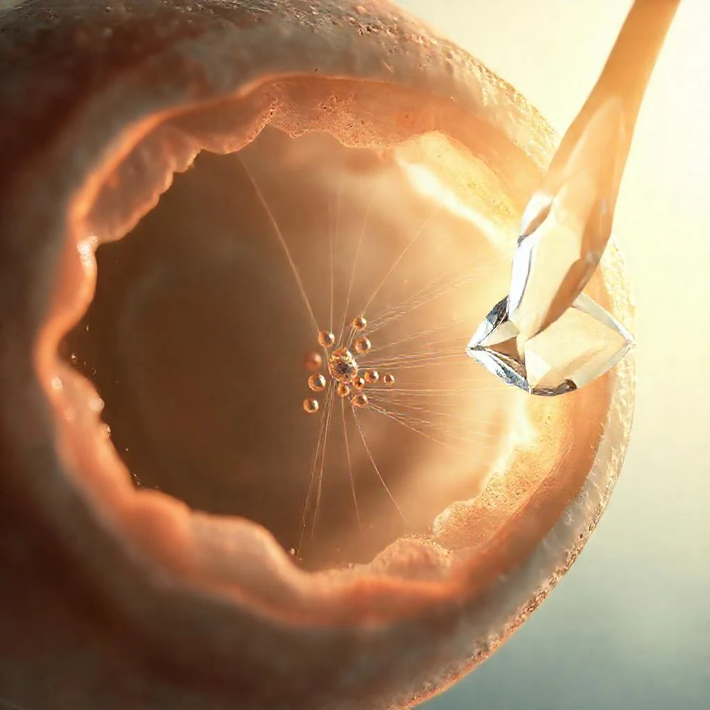
Biologists often tag cells with glowing molecules — it's like searching for a dim light in the smoke. The light quickly fades, and the smoke creates interference. Scientists replaced this approach with a magnetic compass: the cells received tiny magnetized particles, and the signal is read by a diamond sensor with NV centers in a carbon matrix — artificial defects that, like a compass needle, respond sensitively to magnetic fields. The sensor is placed in optical tweezers — a laser beam that holds cells in a stream of liquid without contact — and measures the deflection of the magnetic 'needle'. Even a single tagged cell generates a clear signal of 89 microtesla with noise of just 3.9 — as if a compass picks up the breath of a tiny magnet. Amazingly, the diamond sensor is so sensitive that it can detect the magnetic field of a single electron — no bulky equipment is needed. This 'magnetic eye' paves the way for diagnosing diseases right inside the body, tracking changes in real time.
Diamond NV centers are engineered defects: a nitrogen atom sitting next to a vacancy in the crystal. Their sensitivity can capture the magnetic field of a single electron from several nanometers away.
This precision brings us closer to sci-fi medical scanners — like the tricorder from Star Trek, which diagnoses diseases one cell at a time.