Three Beams Against Cancer: What a Spectroscope Sees in a Drop of Blood express
A drop of blood from a vein falls under three beams, and each one drags its own clues into the light. The infrared beam makes molecules vibrate — from that trembling, you learn how much sugar and fat is in the sample. A green laser scatters off proteins and lipids, shifting color — revealing the remodeling of cell membranes. Ultraviolet light carves out a faint glow from amino acids and vitamins — a metabolic portrait. Instead of hunting for a single mutation, spectroscopy captures hundreds of biochemical fingerprints at once.
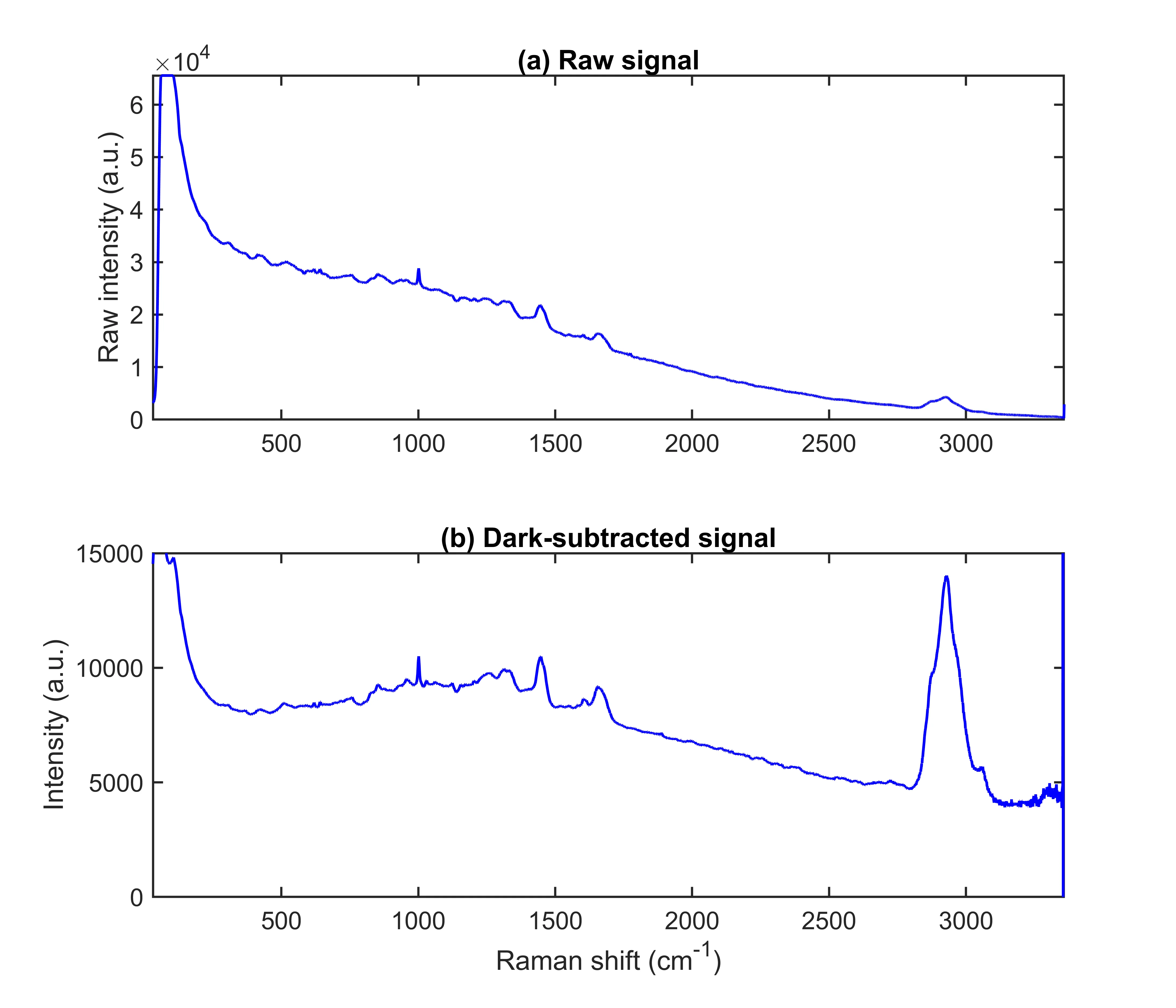
The authors took serum from patients with breast cancer, colorectal cancer, and from healthy people. Each sample was run through three instruments — a Fourier-transform infrared spectrometer, a Raman microscope, and a fluorescence scanner with tunable excitation. Then they stitched all the data into one long string — the so-called low-level data fusion (LLDF)—and trained an XGBoost model to tell sick from healthy. It turned out that individually the methods work well, but together they give a near-perfect result: the area under the error curve reached 0.997 for breast cancer and 0.994 for colorectal cancer.
Three beams, like three witnesses, tell one story, but each in its own language. The infrared spectrometer whispers about sugar chains, the Raman one about lipid shells, the fluorescence one about vitamins and breakdown products. When the readings are stitched together, the picture sharpens, as if three blurry snapshots merge into a crisp photo. Such a union could turn a routine blood test into a tool that spots disease long before the first symptoms appear.
Original abstract
as written by the authors arXiv:2605.13218
Cancer is one of the leading causes of death worldwide, making the development of rapid, minimally invasive, label-free and scalable diagnostic strategies a major challenge in modern oncology. In this context, spectroscopic liquid biopsy has emerged as a promising alternative, as it enables the holistic characterization of biochemical alterations in biological fluids. In this work, we propose a multimodal spectroscopic liquid biopsy framework for multicancer detection based on the combination of Fourier Transform Infrared (FTIR) spectroscopy, Raman spectroscopy, and Excitation-Emission Matrix (EEM) fluorescence spectroscopy together with Machine Learning (ML) methodologies. Serum samples from breast cancer patients, colorectal cancer patients, and healthy controls were analyzed through the three spectroscopic modalities. After modality-specific preprocessing, low-level data fusion (LLDF) was employed to integrate the complementary biochemical information encoded within the different spectroscopic measurements, and classification was performed using XGBoost models. Seven experimental configurations were evaluated, including the three unimodal approaches, all pairwise bimodal configurations, and the full multimodal approach of FTIR, Raman, and EEM fluorescence. The results show that although several individual modalities achieved high discrimination performance, the multimodal fusion provided the most balanced overall results, reaching a ROC-AUC of 0.997 for breast cancer and 0.994 for colorectal cancer, together with highly balanced sensitivity and specificity values.
Serum fluorescence depends on what you ate the day before: riboflavin from eggs and milk glows greenish, while porphyrins from meat glow red. That's why the study strictly controlled participants' diets.
In the novel "Minority Report," criminals were caught before they committed crimes; spectroscopic biopsy promises a future where cancer is found before the tumor.