Enzymes in a Dense Biopolymer: Who Leads Whom ✛
Context
Enzymatic degradation of biopolymers is everywhere, from digestion to converting plant biomass into biofuel. In most applications, the enzyme is applied to one side of a dense matrix, and degradation spreads as a front whose direction is set by the enzyme concentration gradient. Related work in the archive covers the dynamics of biopolymer droplets, molecular transport in cells, and polymer behavior in active media. However, those studies either assume a uniform enzyme distribution or focus on only one aspect: enzyme motion or matrix changes. The link between enzyme penetration and substrate breakdown during directed degradation remained unclear. This work is the first to measure both processes simultaneously with spatial and temporal resolution.
Methods
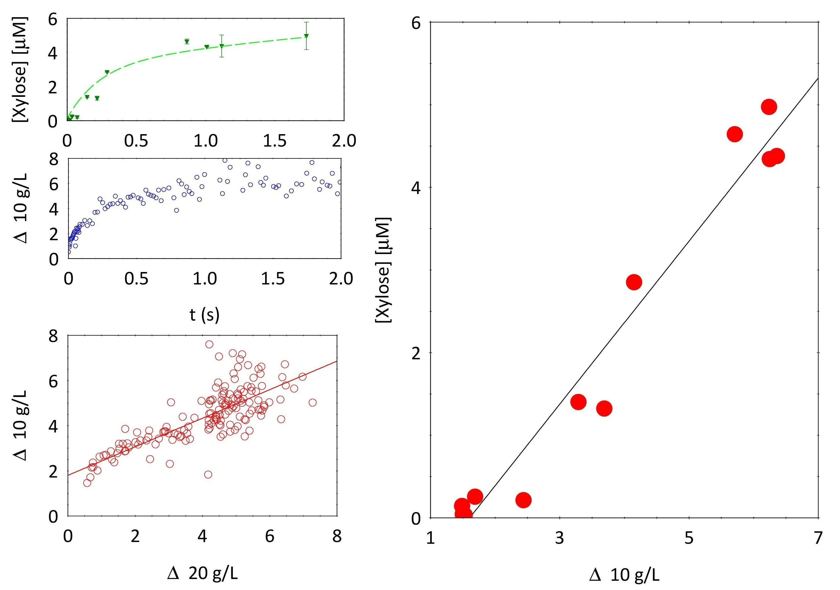
The authors used a model pair: feruloylated arabinoxylan (FAX), a polysaccharide from cereal cell walls, and xylanase, an enzyme that randomly cleaves its chains. A FAX solution at 10 or 20 g/L was placed in a cuvette, and a solution of fluorescently labeled enzyme was gently layered on top. A custom-built setup, Fluo-PCI, combined photon correlation imaging (PCI) and fluorescence microscopy: a laser sheet illuminated the sample, scattered light went to one camera to measure polymer microscopic mobility, and fluorescence went to another to track enzyme concentration. Polymer mobility Δ(z,t) was calculated from the degree of correlation of scattered light, enabling degradation maps in depth–time coordinates. The degradation front was determined by a mobility threshold using Otsu's algorithm. Enzyme diffusion was described by an analytical solution of the one-dimensional diffusion equation for two particle species: large (enzyme) and small (free fluorophore).
Results
The degradation front advanced according to a near-power law: z* ~ (t*)^0.5 for the wild-type enzyme and with an exponent of about 0.7 for a mutant with strongly reduced activity. The front speed increased with decreasing polymer concentration and increasing enzyme concentration. Enzyme diffusion was the same under all conditions: the diffusion coefficient of the large particles was (1.02±0.37)×10⁻¹⁰ m²/s, corresponding to a hydrodynamic radius of about 3.2 nm, consistent with the enzyme size. The small fraction, with a coefficient of (6.59±0.77)×10⁻¹⁰ m²/s, corresponded to free fluorophore. Importantly, diffusion did not depend on enzyme activity: the mutant moved the same as the wild type, ruling out enhanced diffusion effects (EED). A Michaelis–Menten model with spatiotemporal enzyme concentration and an additional activity decay factor f(t) = 0.92·exp(−4.6×10⁻⁵ t) + 0.08·exp(−2.7×10⁻⁶ t) (in seconds) accurately reproduced all experimental mobility maps. For the mutant, a single exponential with a constant of 1.03×10⁻⁵ s⁻¹ sufficed.
Implications
This work stands somewhat apart from its neighbors: those either study polymer dynamics without enzymes or molecular transport in cells without considering matrix degradation. Here, both measurements are combined for the first time in a single experiment, with millimeter spatial resolution and temporal resolution of tens of milliseconds. This allowed separating the contributions of diffusion and reaction kinetics and revealed the key role of slow enzyme deactivation. This approach could become a benchmark for studying directed enzymatic degradation in dense media.
Future development
The Fluo-PCI method is applicable to any enzyme–biopolymer pair where fluorescent labeling is possible. One can investigate the influence of viscosity, pore size, charge, and specific interactions on the coupling between diffusion and degradation. Studying systems with multiple enzymes working synergistically, such as cellulase cocktails for lignocellulose processing, is promising. Extending the model to account for spatial heterogeneity of deactivation and possible product inhibition will allow more accurate predictions of process dynamics in real bioreactors.
Impact
The results directly impact the optimization of enzymatic hydrolysis of biomass for biofuel and bioplastic production, as well as the development of controlled drug delivery systems with enzyme-sensitive carriers.
Next steps
Test the model's universality on other substrates and enzymes, including those with different degrees of polymer network crosslinking. Investigate the mechanism of enzyme deactivation in dense matrices at the molecular level.
Key open problems
In the field of enzymatic degradation of biopolymers, the nature of enhanced enzyme diffusion (EED) remains an open question: it is observed in some systems but not others. This work shows the absence of EED for xylanase in a semi-dilute arabinoxylan solution, adding an argument to the debate. Another unresolved problem is predicting the efficiency of enzyme cocktails in heterogeneous substrates, where local substrate availability and enzyme inactivation vary in space and time.
The knowledge machine's view
This section is addressed to the author of the paper and their colleagues. Neighbouring works were found by meaning-based search across our archive, not by word overlap. We do not review: we show the field around the work.
What the work does
The authors built a setup called Fluo-PCI that simultaneously measures the diffusion of a fluorescently labeled enzyme and the microscopic mobility of polymer chains in a gel. They showed that the degradation front advances according to a power law, while enzyme diffusion is independent of its activity and polymer concentration. A Michaelis–Menten model that accounts for activity decay reproduces the experimental data.
Where its strength is
This work is the first to link enzyme penetration with substrate degradation under directed degradation conditions, providing a quantitative basis for optimizing biotechnological processes.
Where it could go next
- A related study on entropic forces in growing biopolymer droplets is nearby: similar mechanisms might influence local chain mobility near the degradation front.based on: Entropic Frontier: Why Growing Biopolymer Droplets Compress at the Edges arXiv:2606.31786
- It might be interesting to compare the observed front dynamics with models of active media, where polymer networks are rearranged by molecular motors.based on: How Cell Motors Set Droplet Size arXiv:2604.08316
- The archive contains an analysis of solvent effects on the cell wall: transferring that approach to other biopolymer matrices could reveal universal patterns of degradation.based on: How alcohol and chloroform loosen the cell wall arXiv:2605.08780
Worth trying
- Test whether the pore size distribution in the gel changes as the front advances, using methods from the study on pores in membranes.
- Try a model that accounts for heterogeneity of the polymer network, as in the analysis of the entropic front.
- Compare degradation kinetics with cytoskeleton assembly dynamics to identify common principles of biopolymer remodeling.
Why this may matter
Understanding the connection between enzyme transport and matrix degradation is important for the rational design of enzymatic processes in biotechnology, including biomass processing and drug delivery.
Nearby works in our archive
- Proteins bend the membrane the way a spoon bends the film on milksimilarity 0.53
- Entropic Frontier: Why Growing Biopolymer Droplets Compress at the Edgessimilarity 0.51
- How Cells Build a Growth Boundarysimilarity 0.50
- How Cell Motors Set Droplet Sizesimilarity 0.50
- Cosmic filaments pull in two directionssimilarity 0.49
- Pores in the Membrane: Three Keys to Accurate Calculationsimilarity 0.49
- How alcohol and chloroform loosen the cell wallsimilarity 0.48
The archive is sparse around this work: the closest review is only 0.53, against a usual 0.64. It looks like few have drilled nearby.
The exponent of 0.5 in the front motion law for the wild-type enzyme initially suggested purely diffusion-controlled behavior, but data for the mutant with an exponent of 0.7 and modeling showed this to be a coincidence: the front actually slows down due to gradual loss of enzyme activity.
Key numbers
- enzyme diffusion coefficient: 1.02×10⁻¹⁰ m²/s
- enzyme hydrodynamic radius: 3.2 nm
- front exponent for WT: 0.515±0.003
- fast deactivation constant: 4.6×10⁻⁵ s⁻¹
- slow deactivation constant: 2.7×10⁻⁶ s⁻¹