Light Through Skin Reveals Bone Fragility express
A finger is illuminated with a laser beam, and some of the light returns after traveling inside. That's how Raman spectroscopy works: molecules in bone and skin alter the color of the reflected beam differently, leaving their chemical fingerprints. But skin, muscle, and fat are noisy: their signal drowns out the bone's, like a crowd's chatter overpowering a single singer.
To hear that voice, the light was detected not at the entry point but 3 and 6 millimeters away. Light that travels sideways dives deeper into tissue and is more likely to encounter bone. Then a trainable program took over: it was shown thousands of pairs — signals through skin and clean signals from the same bone, now exposed. The program learned to subtract the interference and recover the bone spectrum.
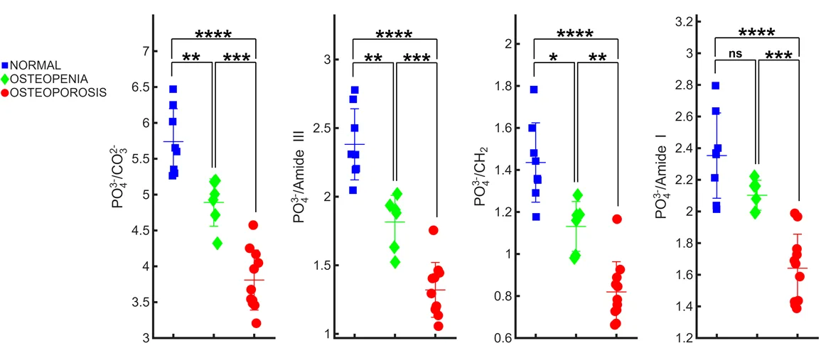
Tests on samples from deceased donors showed that the predicted bone signal distinguishes healthy individuals, those with osteopenia (moderate density loss), and those with osteoporosis. Four metrics computed from the spectrum showed statistically significant differences between groups. Moreover, predictions correlated with standard densitometry results (r = 0.73), approaching the accuracy of direct measurements on exposed bone (r = 0.9).
Initial experiments on two living volunteers confirmed that the same bone features visible in lab samples can be seen through skin. A laser beam passing through a finger could become a simple, safe way to check skeletal strength — no X-rays or bulky machines needed.
Raman spectroscopy is named after Indian physicist Chandrasekhara Venkata Raman, who discovered the effect of light color change upon scattering in 1928 and won the Nobel Prize for it.
In the series 'Star Trek,' the medical tricorder scans a patient without contact and instantly gives a diagnosis — laser bone analysis through skin is a first step toward such a device.