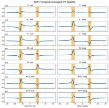
NMR spectroscopy uses the magnetic properties of nuclei. Nuclei with non-zero spin (intrinsic angular momentum) possess a magnetic moment. In an external field, energy levels split (Zeeman effect), and transitions between them can be induced by radiofrequency radiation. The resonant frequency (Larmor frequency) is directly proportional to the field: for example, protons in a 9.4 T field resonate at 400 MHz. But nuclei in molecules are surrounded by electrons, which slightly shield the external field, so the frequency shifts — this is called chemical shift. From these shifts, the chemical environment of the atom is determined. Modern spectrometers obtain a spectrum by emitting a short pulse and recording the decaying signal (free induction decay), which is then converted to a frequency spectrum using Fourier transform.
History
The prerequisites emerged in the 1930s when I. Rabi developed the method of magnetic resonance in molecular beams (Nobel 1944). In 1946, F. Bloch (measured induced electromotive force) and E. Purcell (radiofrequency absorption) simultaneously discovered NMR in solids and liquids, for which they received the Nobel Prize in 1952. In 1966, R. Ernst proposed the pulsed Fourier transform method, revolutionizing sensitivity. Further development of multidimensional spectroscopy (recognized with the 1991 Nobel Prize to Ernst) and stabilization of superconducting magnets pushed resolution to the sub-millihertz level. Today NMR is a standard structural analysis method in chemistry, biology, and medicine (MRI).
How it works
Key Larmor equation: ω = γB₀ (where ω is angular frequency, γ is the gyromagnetic ratio). In liquids, due to rapid molecular motion, interactions are averaged, and narrow lines are observed, with positions characterized by chemical shift: δ = (ν_sample − ν_ref)/ν_ref · 10⁶, measured in parts per million (ppm). Shielding is described by the magnetic shielding tensor, which depends on the local electronic environment. Spin-spin coupling (J-coupling) between nuclei, transmitted through chemical bonds, splits lines into multiplets. A spectrometer consists of a superconducting magnet, radiofrequency generator, signal reception unit, and a computer for accumulation and Fourier transformation of the signal. Pulse sequences (gradient and multiplexed) allow encoding frequencies in multiple dimensions, extracting information about spatial proximity of atoms (nuclear Overhauser effect) and connectivity of the spin system.
In practice
MRI in hospitals is the very same NMR; this way doctors see soft tissues of the body without incisions. Chemists use NMR to determine the structure of new molecules, like a detective reconstructs a scene from clues.
Fun fact
The most sensitive NMR spectrometer can detect a signal from one millionth of a gram of substance. And in brain MRI, signals are collected from a glass of water, but with a resolution that allows discernment of individual neural pathways.
A courtroom interpreter catches every word, every breath, every whisper in the corner. They have no right to miss a detail, because the meaning of the whole conversation depends on it. Quantum state tomography works in much the same way: it reconstructs the full quantum state of a system from a set of measurements, leaving no detail out.
Surf doesn't roll onto the shore as an even front. It comes in tongues of foam that merge and split apart. Magnetic fields inside materials behave much the same way: they breathe in waves, and until now nobody had seen them whole.
An aluminum sphere the size of a large globe sits in a neutron beam and answers each hit with a precise note. Inside the sphere there is nothing but nitrogen and eleven tiny steel balls, each the size of a pinhead. When a neutron enters the sphere, it knocks a particle out of a nitrogen atom, and that jolt turns into an electrical signal.
In the layered crystal CrSBr, the magnetic moments of atoms are arranged like notes in a score: within each layer they point in the same direction, but between layers they point in opposite directions. This antiferromagnetic melody plays up to 132 kelvin, beyond which thermal motion destroys the order.
A tennis ball hits a sandcastle. The castle crumbles, but by where and how far the grains of sand fly, you can guess the castle's shape. That's roughly how the method of exploding proteins with X-rays works. The laser beam is so powerful that the protein molecule loses electrons and flies apart into charged fragments, like the castle under the ball's impact.
Inside an atomic nucleus, protons and neutrons don't sit still like peas in a pod. Each particle occupies its own orbit — a region of space where it can be found. The larger the orbit, the more 'smeared out' the particle is.
To determine the size of the proton cloud in an atomic nucleus, physicists collide an unstable nucleus with a target and observe how many protons it loses. The larger the projectile nucleus, the more often it grazes target nuclei and loses particles.
A protein is like a tiny machine that switches on and off on command. The command arrives when another molecule sticks to it, like a key in a lock. The protein then changes shape and passes the signal along. But exactly how the signal travels inside the protein has long been a mystery.
The instrument shines an infrared beam at a sample, and molecules respond with their own absorption pattern, like bells of different tones. This pattern, the spectrum, holds information about the chemical composition. But to read it, a specialist usually consults a library of reference records, which is slow and not always possible.
When a heavy atomic nucleus splits into two fragments, an invisible quantum thread remains between them. This thread is entanglement: the state of one fragment is instantly linked to the state of the other, as if they were still a single whole. For the first time, physicists have precisely calculated how long and strong such a thread can be, tracing the fission process step by step.
The atomic nucleus is usually pictured as a dense ball where protons and neutrons are mixed together. But in some isotopes, neutrons can stick out far beyond this ball, forming a diffuse shell — a neutron halo. This is especially common in nuclei with many more neutrons than protons.
The atomic nucleus isn't always round like a billiard ball. Some nuclei stretch out like a rugby ball, while others become pear-shaped — with one end thicker than the other.
A uranium-235 nucleus that captures a slow neutron doesn't immediately fly apart into fragments. Instead, it seems to get stuck in two 'pits'—states with different shapes. The first, the ground state, is a slightly elongated drop. The second, the isomeric state, is a drop stretched further but not yet torn.
The pendulum in these clocks doesn't swing from a spring or a weight, but from a tiny kick inside the atomic nucleus. Ordinary atomic clocks keep time by the oscillations of electrons, but here the pendulum is the nucleus itself — a dense ball of protons and neutrons.
Boiling milk develops a trembling skin. By watching its movements, you can guess the strength of the fire beneath the pot and the fat content of the milk. In a similar way, astrophysicists peer into the ancient microwave radiation — the oldest light that has reached us from an era when the universe was only 380,000 years old.
A radio receiver the size of a palm can measure signal power more accurately than expensive lab benches. Inside, there are no coils or antennas, just a glass head and a cloud of rubidium atoms cooled to near absolute zero.
Ultra-sensitive scales try to detect the difference between two nearly identical grains of sand. One is a perfect mirror reflection of the other. In the world of elementary particles, such mirror reflection is called P-parity, and its violation is one of the great mysteries of physics.
Inside the atomic nucleus, protons and neutrons are packed so tightly that the forces between them resemble tightly coiled springs. Some springs compress and expand equally in all directions: these are the familiar central forces.
High in the mountains, where the air is clear and still, physicists are preparing to catch elusive visitors from space — neutrinos. These particles, like ghosts, pass straight through the Earth, leaving almost no trace. But occasionally a neutrino does slam into an atom, producing a shower of particles that can be seen.
X-ray magnetic circular dichroism is a precise tool that has long allowed the separation of spin and orbital magnetic moments. However, the classical approach resembles judging an orchestra by its volume: it relies on sum rules, reducing the entire fine structure of the spectrum to a couple of integrals.
In brachytherapy, thin catheters are inserted into tumors. On CT scans, they look like tangled glowing threads. Previously, doctors would manually outline each one, spending up to an hour.
The new algorithm learns from synthetic scans — drawing metal strips in an empty volume, like threads without a body.
In the first seconds after the Big Bang, the Universe left us not only light but also something far more elusive — the relic neutrino background. This echo of creation permeates every cubic centimeter, yet we barely notice it.
For almost a century, seismology has been the planet’s chief X-ray, probing the interior through elastic waves. But neutrinos, ghost particles predicted by Wolfgang Pauli, offer a different perspective.
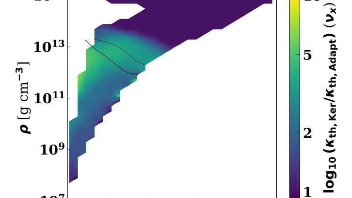
The collision of two neutron stars is a cosmic laboratory where matter is compressed to unimaginable densities. In the first moments after the merger, a hot cloud forms and cools by emitting neutrinos. These ghostly particles carry away energy and dictate how much matter is ejected into space and which heavy elements are synthesized.
Deep underground, iron glows white-hot, and the pressure is so intense that an ordinary instrument would be crushed flat. To peer into this world, physicists built a special microscope based on tiny diamond defects (so-called NV centers).
When a building collapses, steel rebar turns into a chaotic magnetic compass. Entropy, a measure of disorder, increases, but it's this very disorder that points to voids where people might be.
In a small Petri dish sits a droplet of liquid the size of a pinhead. An ordinary scanner wouldn't even notice it: the signal is too weak. But bring a tiny magnetic field sensor close to the droplet, and the picture changes.
Trying to count people in a dark room by the sound of their breathing hits a wall of background noise — individual breaths are inaudible. Scientists working with quantum spins in solids face a similar challenge. Each spin (the electron's magnetic moment) has its own "voice," but it is drowned out by the technical noise of the instruments.
Atomic nuclei are rarely perfect spheres. More often, they resemble a slightly squashed ball — the electric charge inside is unevenly distributed, and protons may shift a bit, creating a tiny 'pear-shaped' bulge.
A pendulum that almost nothing can knock off balance (not shaking, not heat, not magnetic fields) because it is hidden in the very heart of the atom. That is the kind of pendulum physicists used when they first switched on a nuclear clock. Previously, the best clocks, optical ones, relied on electron transitions.
An old photograph taken at the moment of the universe's birth has been carrying its light to us for nearly 14 billion years. This snapshot is the cosmic microwave background, the cooled echo of the Big Bang. It captures the tiny inhomogeneities from which galaxies later grew.
Magnetic particles are attached to the surface of the diamond by DNA strands, like puppets on strings. They constantly dance under the thermal motion of the liquid molecules. The liquid's viscosity sets the pace: in honey, the particles barely tremble; in alcohol, they dance quickly.
The atomic nucleus rarely sits still: it often trembles, rotates, or pulses like a plucked string. Each such motion is an excited state, and describing it usually requires extremely complex calculations. Now neural networks come to the rescue: they shape the nucleus the way a musician identifies a note in a chord by ear.
Many chemical transformations resemble assembling furniture in complete darkness: you see the starting parts and the finished cabinet, but the assembly itself remains a mystery. The intermediate states, like awkward positions of the craftsman's hands, vanish too quickly to be discerned.
The atomic nucleus is a natural spring of colossal power. When the protons and neutrons inside it rearrange, it enters a special long-lived state — a nuclear isomer. It stores energy like a spring compressed to its limit.
Two ultra-dense stars spiral around each other and eventually collide. This dance lasts only moments, but it sends powerful ripples through space — gravitational waves. After the merger, a rapidly spinning blob remains, ringing like a bell, and that ring carries information about what the stars were made of.
A spectrum is like a photograph of a material's inner workings, taken with light. When a beam passes through or reflects off a sample, molecules leave characteristic dark bands, peaks, on that photograph. But if the image is overexposed or veiled in gray, making out details becomes nearly impossible.
We know about neutron stars thanks to the work of Fritz Zwicky, Subrahmanyan Chandrasekhar, and Jocelyn Bell Burnell. They are former cores, squeezed to city size: a teaspoon of their matter weighs as much as Mount Everest.
The Standard Model is a set of rules for all known particles. But it says nothing about dark matter, which makes up a quarter of the Universe. To find it, you don't need to build city-sized colliders. Radioactive molecules act like sniffer dogs: their incredible 'scent' picks up the faintest traces of unknown forces that slip past giant machines.
In a benzene molecule, electrons buzz around the nuclei like a swarm of bees around a hive. But this swarm doesn't just hum: it pulses: every 856 attoseconds, the electron cloud completes a full oscillation cycle. The hive itself, the carbon nuclei, rocks more slowly, with a period of 36 femtoseconds.