Intercalation Memory in MXenes: How Layers Remember Their History
Context
The work sits at the intersection of intercalation research in layered materials and single-nanoparticle microscopy. Neighboring studies explore viscous films near mineral walls, an oxygen key to superconductivity, oxide membranes with memory, twisted crystals, enzymes in gels, exciton condensates in graphene, X-ray crystallography without guesswork, and optical tweezers. In this lineup, the present paper stands out by being the first to link nanoscale heterogeneity in layer stacking to macroscopic electrochemical memory, using direct optical monitoring of individual flakes.
Methods
The scientists built a custom microscope combining interferometric reflectance microscopy and interferometric scattering, with time resolution down to microseconds and localization accuracy better than 10 nm. Ti3C2Tx samples were deposited on indium tin oxide-coated glass and placed in an electrochemical cell with sulfuric acid. Optical contrast was calibrated against atomic force microscopy to determine layer number. In parallel, they performed cyclic voltammetry and synchrotron X-ray diffraction on a film about 3 μm thick.
Results
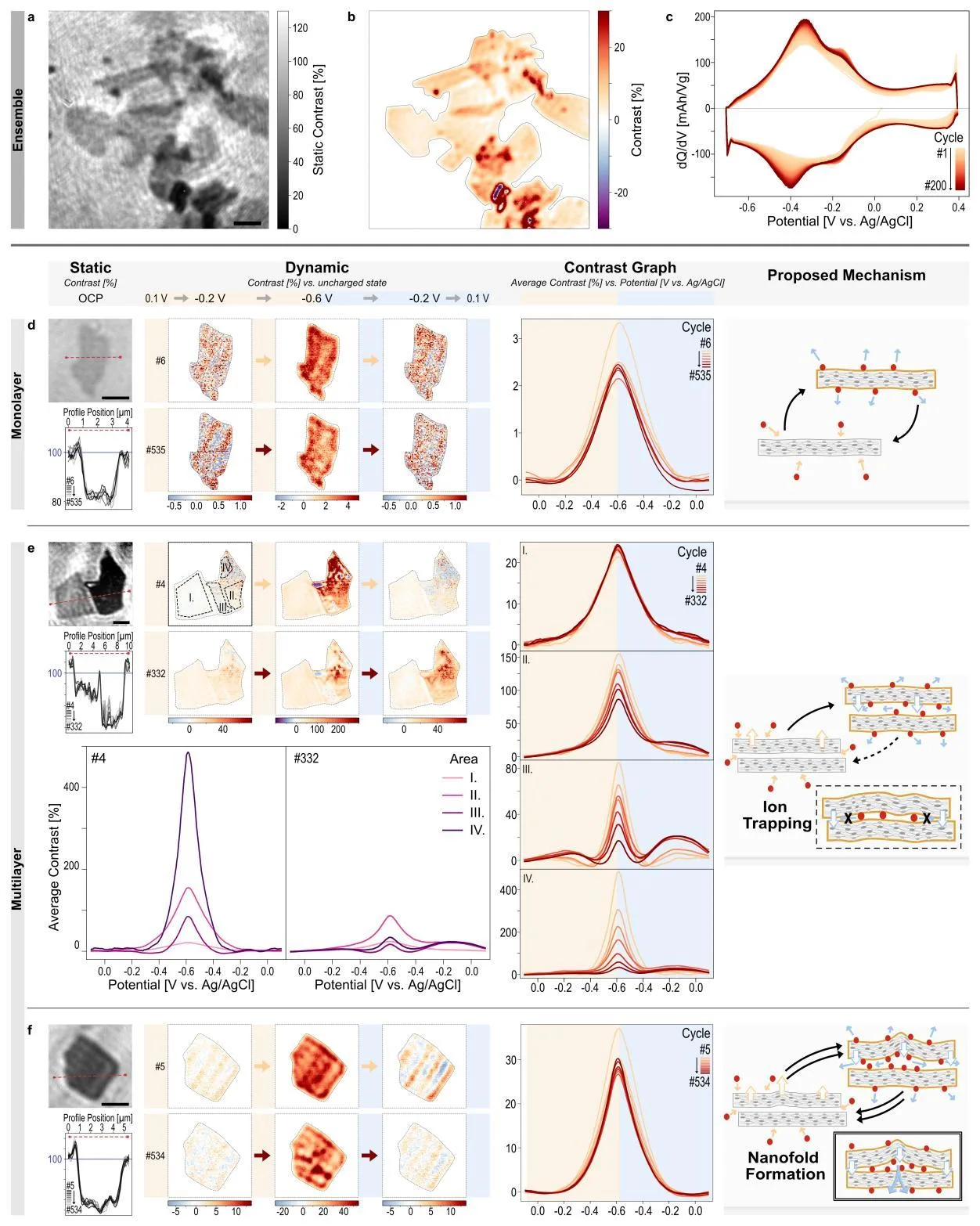
Monolayer Ti3C2Tx shows nearly perfectly reversible response: the contrast peak drops by only 2% when the scan rate increases from 10 to 100 mV/s. For six or more layers, the drop reaches 36%, and the b-value decreases from 0.978 to 0.711, indicating a transition from surface-controlled to diffusion-controlled kinetics. Repeated cycling revealed three scenarios: monolayers remain stable, restacked multilayers accumulate residual strain and lose reversibility, while coherently stacked multilayers form reversible folds that completely disappear when the potential returns. Synchrotron diffraction confirmed that the interlayer spacing in the film converges to a new, less breathing state, and the film texture reorganizes within the first few tens of cycles.
Implications
Unlike neighboring studies that examine either averaged ensembles or individual molecules in biological systems, this work shows for the first time how local stacking of a few layers dictates intercalation kinetics and memory. It is the first direct visualization that electrode activation is not a uniform opening of galleries, but a history-dependent reorganization with selection of preferred deformation modes. The results connect nanoscale mechanics to macroscopic electrochemical response, which was previously inaccessible.
Future development
The topic could evolve toward mapping local mechanical stresses and chemical composition inside individual folds using spectroscopy. It may be possible to create libraries of stackings with designed memory for controlling ion transport. Applying the method to other layered materials, such as transition metal dichalcogenides or layered oxides, is promising to determine how universal the folding mechanism is.
Impact
The developed approach is applicable to diagnostics of batteries, supercapacitors, and electrochromic devices, where intercalation determines lifetime. Understanding the role of local stacking will help improve the synthesis of electrodes with controlled morphology.
Next steps
The next step could be combining optical microscopy with local Raman spectroscopy or X-ray probing for chemical identification of states within folds. It is also important to test the behavior of other ions, such as lithium or sodium, and their influence on structural memory.
Key open problems
In materials science, an open question remains how nanoscale heterogeneity determines macroscopic electrode degradation. This work directly links the unresolved problem of early activation and hysteresis in layered materials to specific mechanical events at the level of individual flakes, offering a new path for predicting service life.
The optical contrast of a single Ti3C2Tx monolayer is so small that its change upon intercalation is equivalent to a thickness change of only a few angstroms, comparable to the size of a single hydrogen atom.
Key numbers
- contrast drop for 1L at 10→100 mV/s: 2%
- contrast drop for 6L+ at 10→100 mV/s: 36%
- b-value for 1L (cathodic): 0.978
- b-value for 6L+ (cathodic): 0.711
- film thickness for GIXRD: 3 μm