Scientist

Francois Englert

род. 1932 · Theoretical physics, elementary particle physics
Belgian theoretical physicist, one of the discoverers of the Brout–Englert–Higgs mechanism, which explains how elementary particles acquire mass. He shared the Nobel Prize with Peter Higgs.

Biography

Born in 1932 in Etterbeek, Belgium. He studied at the Free University of Brussels, where he received his doctorate in 1959. He worked at Cornell University, then returned to Brussels, where he became a professor. In 1964, together with Robert Brout, independently of Higgs, he proposed the mechanism of spontaneous symmetry breaking in gauge theories. He won the Nobel Prize in Physics in 2013.

Key discoveries

💡 Englert was almost unknown to the general public before the Nobel Prize; when he received the call from Stockholm in 2013, the first thing he did was send a fax to his co-author Brout, who had passed away in 2011, writing: 'Dear Robert, we did it.' Englert also enjoys painting and playing the cello.
"Brout and he viewed their work on the problem as an attempt to understand how nature could be both symmetric and broken, without consideration for fame."
Concepts
Higgs bosonQuantum FieldStandard Model
Scientists
Satyendra Nath BoseEmilio Gino SegrèPaul DiracPeter HiggsRichard FeynmanJ. J. ThomsonWerner HeisenbergMarie Curie

Related articles

q-bio.QM2026-01-101 minarXiv:2601.06360express
Prostate Cancer: A Look at the Tumor's Surroundings
A glass of water and a glass of jelly look the same but behave differently. So it is with prostate tissue: on a standard slide under a microscope, you can see both the tumor glands and the supporting connective tissue around them. Previously, pathologists focused only on the glands, treating the support as mere background.
Medical Physics2026-02-042 minarXiv:2602.04461express
Light instead of radiation: seeing an organ without a needle
Radioactive tracers are like a courier with a beacon: you inject it into the body, and it highlights the target organ on the scan. But this courier comes with strict rules: you can't send it often, it quickly 'gets tired,' and it costs a lot. Doctors have long dreamed of a way to see an organ without radiation.
Medical Physics2026-02-091 minarXiv:2602.08918express
The Living Lens: How MRI Sees Muscles in Motion
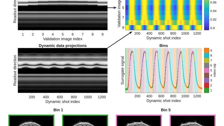
A photographer snaps a runner: to avoid a blurry shot, a short shutter speed is needed. An MRI scanner is like that photographer, but its 'shutter speed' is long — by the time it captures, the muscle has already changed. That's why dynamic MRI (a series of volumetric frames in motion) is a tough challenge: data are scarce, and motion doesn't wait.
HEP Experiment2026-02-191 minarXiv:2602.17165express
Eight collisions that revealed a new process
In the ATLAS detector at the Large Hadron Collider, protons collide billions of times per second. From this stream, physicists fished out eight events in which the collision produced two Z particles and one photon.
q-bio.OT2026-03-011 minarXiv:2603.01054express
How a Bladder Turns into a Pear
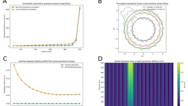
The bladder is like a balloon: as it fills, the wall stretches and the shape changes. Doctors usually monitor the pressure inside and how much fluid has entered, but they rarely look at the geometry itself — how the surface curves. Yet irregularities can appear before the usual metrics deteriorate.
An MRI That Fits in a Backpack
A standard MRI machine is a huge magnet that you can't move, plus a powerful computer that turns signals from the body into a picture. In recent years, portable scanners with weaker fields have appeared: they are light, cheap, and can work in a field hospital or an ambulance.
Medical Physics2026-03-101 minarXiv:2603.09898express
A Scanner That Sees Through Tumors
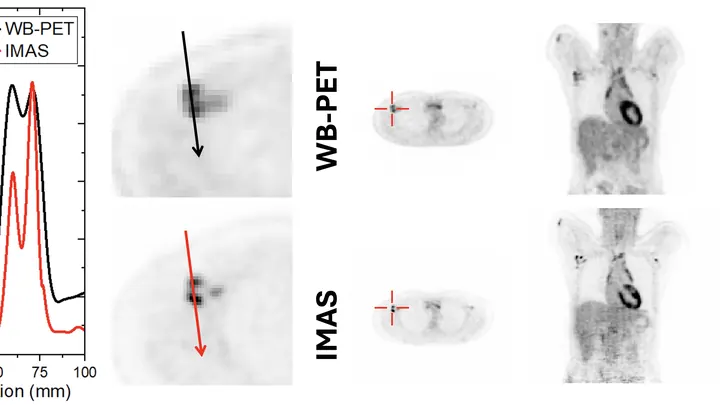
A standard PET scanner is like a camera with a narrow lens: to capture the whole body, you have to move it back and forth and take many shots. The new IMAS tomograph is a wide-angle camera that covers 71 centimeters of the body at once. The patient lies still, and the machine collects an image from head to thighs in a single pass.
Medical Physics2026-03-191 minarXiv:2603.19368express
The Shadow of Cancer on a Clean Scan
PET/CT is like a map where cancer cells light up as bright spots. But what if the map is blank and the disease is still hiding somewhere? In 132 men with recurrent prostate cancer, doctors found no glowing lesions, but they decided to study the texture of the scan itself, like an experienced tracker noticing bent grass where an animal has passed.
Medical Physics2026-03-231 minarXiv:2603.22092express
The Placenta Under the Microscope: How Oxygen Travels to the Fetus
The placenta is like a bustling market where oxygen passes from mother to child. If the stalls are neatly arranged and goods are delivered quickly, everyone gets enough. But when the aisles get clogged, traffic jams start: some places are crowded, others empty.
Medical Physics2026-03-241 minarXiv:2603.22666express
Tracking Prostate Cancer Metastases in Scans
When treating prostate cancer, doctors often see not one tumor but many lesions in bones and lymph nodes. Each lesion can respond to therapy differently: one shrinks, another stays the same, a third grows. Previously, the disease was assessed as a whole, like judging the weather by the average temperature over a year.
q-bio.BM2026-03-241 minarXiv:2603.23775express
Diagnostics in the Palm of Your Hand: Electricity Instead of Optics
In a hospital, blood from a finger prick usually goes to the lab, and the result comes back the next day. The new method promises to cut that wait to a few minutes. Instead of catching light reflected off a sample, it reads the electrical voltage that arises at the boundary between two media, like the faint tingle you feel when you touch a battery with wet fingers.
Medical Physics2026-04-021 minarXiv:2604.01710express
Quiet MRI Machines Just Got Louder
A weak magnet in an MRI machine is like a dim bulb in a dark room: you get a picture, but details drown in grain. These scanners operate at fields below 0.1 tesla (a typical hospital machine runs at 3 tesla, 30 times stronger), yet they're lightweight, inexpensive, and don't require a special room. The catch: noise eats away at quality.
Medical Physics2026-04-112 minarXiv:2604.10227express
Dose Under Control
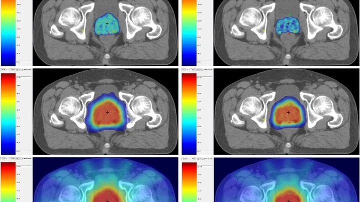
When a doctor irradiates a tumor, they try to spare nearby healthy organs. In the past, the standard approach was to give many small doses of radiation, like watering a lawn with a fine-spray watering can. Now, hypofractionation is increasingly used: fewer sessions, but larger doses, like a high-pressure hose. The question is how to tell whether the neighboring garden beds got overwatered.
Medical Physics2026-05-092 minarXiv:2605.08957express
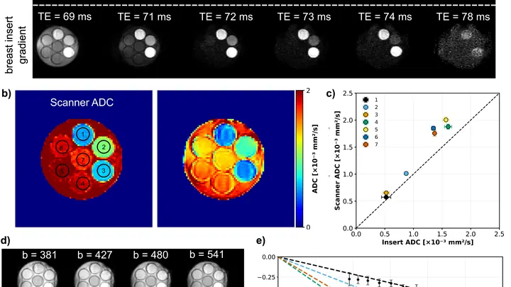
Gradient Coil for the Breast: MRI Without Contrast
A flashlight spreads light in a broad beam, while a laser cuts a narrow path and picks out the finest details. In magnetic resonance imaging (MRI), the gradient, a rapid change in the magnetic field along the body, plays the role of that laser.
Medical Physics2026-05-091 minarXiv:2605.09091express
How MRI is learning to tell water in muscles from water in blood vessels
To peek inside a muscle, it is illuminated with radio waves in a strong magnetic field: that's how MRI works. Water in cells and water in blood respond differently, but a standard scanner doesn't tell them apart: it produces an averaged image where the signal from capillaries is superimposed on the signal from muscle fibers.
Medical Physics2026-05-242 minarXiv:2605.24815express
How to Make MRI See a Beating Heart
A photographer shoots a runner with a long exposure and gets a blurry silhouette. That's roughly how a standard MRI scanner "sees" moving organs: the heart contracts, the lungs breathe, and the image smears. To avoid this, patients are asked to freeze and hold their breath. But for a child or someone short of breath, that's nearly impossible. The method called GRASP tackles the problem differently.
Medical Physics2026-06-062 minarXiv:2606.08065express
Breathing vs. Snapshot
As a person breathes, the liver glides smoothly up and down. When treating a tumor with radiation, this motion must be accounted for: the beam must hit the target, not healthy tissue. Usually, breathing is "captured" using special computed tomography (CT), taking a series of frames over one breath cycle.
Image Processing2026-07-131 minarXiv:2607.11385express
How Cleaning MRI Scans Helps Find Prostate Cancer
Prostate MRI scans often look like photos taken through a dirty window: noise, blurred edges, curvature from field imperfections. Scientists checked what happens if you 'wash' that window: remove the excess, correct the distortions, and accurately align different types of scans.
Applied Physics2026-08-101 minarXiv:2608.10225express
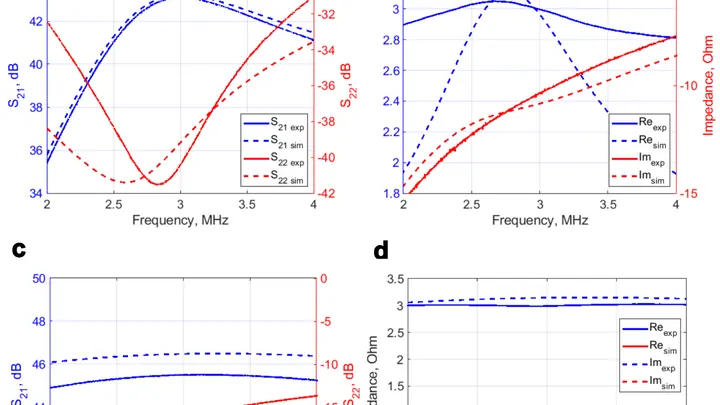
A Quiet Amplifier for a Weak Signal
The coil in an MRI picks up radio signals from hydrogen atoms, like an antenna catching a distant radio station. But the signal is weak, and if the amplifier itself is noisy, the image comes out grainy.