Nuclearmagnetic resonance (NMR) is a physical phenomenon in which atomic nuclei in a magnetic field absorb and re-emit electromagnetic radiation at a characteristic frequency. This frequency depends on the strength of the magnetic field and the magnetic properties of the nucleus, as well as its chemical environment. NMR is a powerful tool for determining the structure of molecules, studying dynamic processes, and imaging the inside of objects. It is the underlying principle behind magnetic resonance imaging (MRI), a widely used medical diagnostic technique.
History
The phenomenon was first described by Isidor Isaac Rabi in 1938, who measured the magnetic moments of nuclei in molecular beams. In 1946, Felix Bloch and Edward Mills Purcell independently developed NMR for condensed matter, for which they shared the Nobel Prize in Physics in 1952.
How it works
When a sample is placed in a strong magnetic field, the nuclear spins align either parallel or antiparallel to the field, creating a small net magnetization. A radiofrequency pulse at the Larmor frequency (the resonance frequency) tips this magnetization into the transverse plane. As the spins precess and relax back to equilibrium, they emit a radiofrequency signal that is detected. The frequency and decay of this signal provide information about the chemical environment and interactions of the nuclei, which can be analyzed to reconstruct structural and dynamic details.
In practice
NMR is used in chemistry and biochemistry for structure determination of molecules, in medicine for MRI imaging, and in materials science for analyzing the composition and properties of materials. It is also used in the oil industry for well logging and in quality control for food and pharmaceuticals.
Fun fact
The first MRI image of a human body was produced in 1977 by Raymond Damadian, who scanned his own chest over a period of nearly five hours.
A sculptor looks at a block of marble and sees the figure within, waiting to be freed from excess material. Instead of adding clay, he removes everything that is not part of the form. The authors of a new study on magnetic resonance took a similar approach: rather than complicating the tissue model by adding ever more details, they stripped away the superfluous to reveal the essence.
The atomic nucleus is usually pictured as a dense ball where protons and neutrons are mixed together. But in some isotopes, neutrons can stick out far beyond this ball, forming a diffuse shell — a neutron halo. This is especially common in nuclei with many more neutrons than protons.
An X-ray flash tears through matter in billionths of a second. To remember it, physicists make atoms briefly absorb the light and then release it back. But pulling out a photon on command is like grabbing the right book from a shelf when the shelves move on their own.
The usual approach is to move several absorbers with extreme precision, like a well-coordinated orchestra.
Fast radio bursts — millisecond radio pulses arriving from distant galaxies — long remained a puzzle. Their colossal energy, compressed into instants, pointed to cataclysms on neutron stars, perhaps pulsars or magnetars.
The enigma of fast radio bursts (FRBs) is not just a question of 'what exploded?' It’s a drama of energy transfer under extreme conditions, where the slightest stumble leads to collapse.
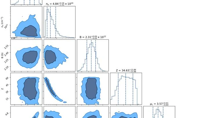
A lighthouse sends its beam through a storm of its own mirrors: that's roughly how fast radio bursts behave when trying to leave a magnetar's vicinity. A magnetar is a neutron star with a magnetic field a quadrillion times stronger than Earth's.
Two ultra-dense stars spiral around each other and eventually collide. This dance lasts only moments, but it sends powerful ripples through space — gravitational waves. After the merger, a rapidly spinning blob remains, ringing like a bell, and that ring carries information about what the stars were made of.
A spinning top whirls so fast it's about to fly apart. Add to it a super-strong magnet — one that, if placed on the Moon, would wipe data from every credit card on Earth. That's a magnetar: an ultra-dense remnant of an exploded star, a lump of matter where a teaspoon weighs as much as a mountain.
A substance's spectrum is its fingerprint. Usually, to record one, you place the sample in a strong magnetic field, like in an MRI scanner. But there is a way to do without a magnet altogether: detect the signal from atomic nuclei at zero field. Until now, this method suffered from poor sensitivity, and its complex spectra were hard to predict.
The crust of a neutron star is not just a solid shell. Under pressures of billions of tons per square centimeter, atoms lose their electrons, and the remaining nuclei arrange into a rigid crystal lattice, reminiscent of a honeycomb, but made of matter a trillion times denser than steel.
Inside the Sun, invisible nuclear transformations occur every second. One of them is the birth of boron-8: a beryllium-7 nucleus captures a passing proton and momentarily becomes a new element. This step kicks off a chain that ends with the emission of a neutrino — a particle capable of passing straight through Earth.
The atomic nucleus is not just a ball of protons and neutrons. Inside it, like a tiny magnet, there is its own distribution of magnetization. Until now, it has been difficult to see: nuclei live for tiny fractions of a second, and their magnetic field is weak. Now physicists at CERN have found a way to look inside.
In particle scattering experiments, the target is often polarized: nuclei are aligned so that their spins point predominantly in one direction. The degree of this alignment affects the collision outcomes.
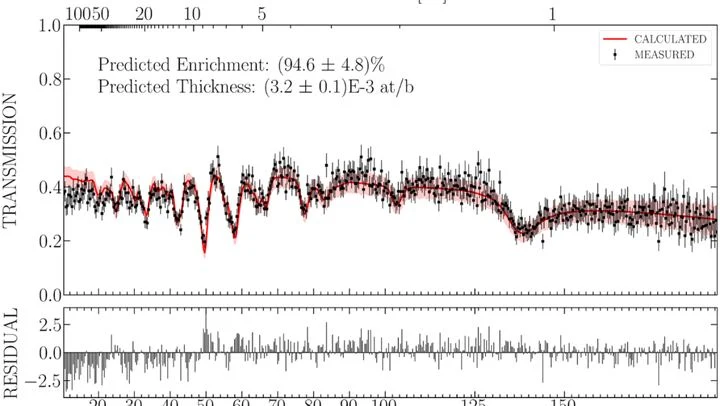
A customs officer can't take a suitcase apart to see what's inside. They shine X-rays through it and, from the shadows on the screen, tell: here's metal, here's organic matter. In much the same way, an inspector for future nuclear disarmament treaties should be able to check a warhead — without opening it or seeing its secret internals.
Neutrons are better suited for this than X-rays.
Scientists peered into a two-dimensional magnet made of nickel atoms arranged in triangles using a method created by Isidor Isaac Rabi — nuclear magnetic resonance (similar to MRI). Inside, they found magnetic arrows that, in a strong field, froze into an 'up-up-down' pattern, like a formation of soldiers: two steps forward, one step back.
A pebble from a distant asteroid can remember the magnetic field that acted on it billions of years ago. Such pebbles, particles from asteroid Ryugu, were brought to Earth by Japan's Hayabusa2 probe. Inside them, like the needle of an ancient compass, the direction of the magnetic field from the solar system's youth is frozen.
A graphene ribbon nine atoms wide is like a track where electrons race, as water rushes down a narrow trough. Gamma radiation is a stream of particles with enormous energy, capable of punching through metal and plastic.
Magnetic resonance is like a tuning fork that rings only at its own note. Each molecule has its own frequency, and from that you can tell what the molecule is and what state it's in. But previously, this tuning fork was listened to through a thick wall: the signal was blurred, and you couldn't make out the exact note.
Every cell is a miniature tuning fork. A sharp magnetic pulse makes it 'ring', and the decay of that sound depends on what's inside: viscosity, density, composition. A tiny diamond with a structural defect acts like a hypersensitive microphone, picking up magnetic echoes from individual cells.